
Be an expert among experts, join us at ODAC to hear from the esteemed faculty members featured in top media outlets.
April 2020
ODAC Launches New Blog
First and foremost, in light of the rapidly evolving global COVID-19 situation, we hope that you and your family are safe and healthy. We send companywide thoughts and prayers to the individuals, families, and other groups who have been impacted by this situation, and hope that things will improve very soon.
While many things are uncertain surrounding this virus, as an organization, SanovaWorks believes in taking action early. We are dedicated to providing the best care and support we can to all our employees and business partners.
We have the extremely good fortune to be functioning already as a virtual company and because of this we hope to be able to provide support and resources to our entire network who might not have the experience we have. Please check our blog for our tips and recommendations for transitioning to and being successful in a remote work environment:https://sanovaworks.com/2020/03/11/top-immediate-needs-of-remote-employees/
In addition to this, in order to protect our teams and others, until further notice, we have issued a complete restriction on all business-related travel. While the CDC has not placed restrictions on domestic travel, they have recently posted travel warnings on their website: https://www.cdc.gov/coronavirus/2019-ncov/travelers/travel-in-the-us.html?mod=article_inline
The CDC also provide general recommendations that we should all be following to prevent the spread of this disease:https://www.cdc.gov/coronavirus/2019-ncov/about/prevention.html
We will be working diligently as teams to connect with many of you so that we can share some very interesting ways to accomplish our results in this new, virtual environment. We have many years of experience transitioning traditional programs to digital programs, and launching successful virtual programs. Because we are already positioned as a remote company that produces virtual programs, we hope that we are able to support your own initiatives and bridge the gap this global situation has caused.
And last but not least, with a shout out to Jim Collins who introduced me to the Stockdale Paradox in his book Good To Great, we all need to look squarely at the facts, but have confidence that together we will prevail, as we balance realism with optimism.
Together.
Shelley and the entire SanovaWorks Team
March 2020
Statement From Our President and CEO Regarding COVID-19
First and foremost, in light of the rapidly evolving global COVID-19 situation, we hope that you and your family are safe and healthy. We send companywide thoughts and prayers to the individuals, families, and other groups who have been impacted by this situation, and hope that things will improve very soon.
While many things are uncertain surrounding this virus, as an organization, SanovaWorks believes in taking action early. We are dedicated to providing the best care and support we can to all our employees and business partners.
We have the extremely good fortune to be functioning already as a virtual company and because of this we hope to be able to provide support and resources to our entire network who might not have the experience we have. Please check our blog for our tips and recommendations for transitioning to and being successful in a remote work environment:https://sanovaworks.com/2020/03/11/top-immediate-needs-of-remote-employees/
In addition to this, in order to protect our teams and others, until further notice, we have issued a complete restriction on all business-related travel. While the CDC has not placed restrictions on domestic travel, they have recently posted travel warnings on their website: https://www.cdc.gov/coronavirus/2019-ncov/travelers/travel-in-the-us.html?mod=article_inline
The CDC also provide general recommendations that we should all be following to prevent the spread of this disease:https://www.cdc.gov/coronavirus/2019-ncov/about/prevention.html
We will be working diligently as teams to connect with many of you so that we can share some very interesting ways to accomplish our results in this new, virtual environment. We have many years of experience transitioning traditional programs to digital programs, and launching successful virtual programs. Because we are already positioned as a remote company that produces virtual programs, we hope that we are able to support your own initiatives and bridge the gap this global situation has caused.
And last but not least, with a shout out to Jim Collins who introduced me to the Stockdale Paradox in his book Good To Great, we all need to look squarely at the facts, but have confidence that together we will prevail, as we balance realism with optimism.
Together.
Shelley and the entire SanovaWorks Team
March 2020
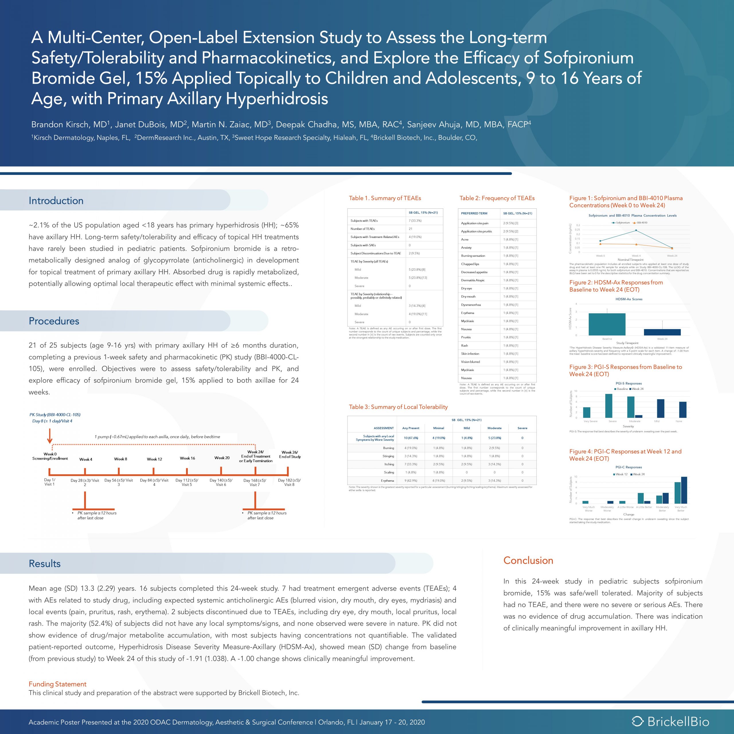
Long Term Use of Novel Therapeutic for Topical Treatment of Primary Axillary Hyperhidrosis in Pediatric Subjects
Source: ODAC Dermatology, Aesthetic & Surgical Conference (ODAC) Discovery in Dermatology Poster Session
At the 17th Annual ODAC Dermatology, Aesthetic, and Surgical Conference (ODAC) held January 17-20 in Orlando, FL, Brandon Kirsch, MD, Janet DuBois, MD, Martin N. Zaiac, MD and Deepak Chadha, MS, MBA, RAC presented scientific research of long term data with a novel therapeutic for topical treatment of primary axillary hyperhidrosis in pediatric subjects.
Discovery in Dermatology
The use of retro-metabolically designed drugs in dermatology is novel and has the potential for providing significant therapeutic benefit to pediatric and adult patients.
Sofpironium bromide is an ester analogue of glycopyrrolate that inhibits muscarinic receptors in sweat glands. It was developed according to the principles of retro-metabolic drug design, in which the goal is to create an active compound that is metabolized in vivo to an inactive moiety in a single, predictable reaction. Retro-metabolically designed drugs are rapidly metabolized in the bloodstream, potentially allowing for optimal therapeutic effect at application sites with minimal systemic side effects.
Click to enlarge
Introduction
~2.1% of the US population aged <18 years has primary hyperhidrosis (HH); ~65% have axillary HH. Long-term safety/tolerability and efficacy of topical HH treatments have rarely been studied in pediatric patients. Sofpironium bromide is a retro-metabolically designed analog of glycopyrrolate (anticholinergic) in development for topical treatment of primary axillary HH. Absorbed drug is rapidly metabolized, potentially allowing optimal local therapeutic effect with minimal systemic effects..
Procedures
21 of 25 subjects (age 9-16 yrs) with primary axillary HH of ≥6 months duration, completing a previous 1-week safety and pharmacokinetic (PK) study (BBI-4000-CL-105), were enrolled. Objectives were to assess safety/tolerability and PK, and explore efficacy of sofpironium bromide gel, 15% applied to both axillae for 24 weeks.
Results
Mean age (SD) 13.3 (2.29) years. 16 subjects completed this 24-week study. 7 had treatment emergent adverse events (TEAEs); 4 with AEs related to study drug, including expected systemic anticholinergic AEs (blurred vision, dry mouth, dry eyes, mydriasis) and local events (pain, pruritus, rash, erythema). 2 subjects discontinued due to TEAEs, including dry eye, dry mouth, local pruritus, local rash. The majority (52.4%) of subjects did not have any local symptoms/signs, and none observed were severe in nature. PK did not show evidence of drug/major metabolite accumulation, with most subjects having concentrations not quantifiable. The validated patient-reported outcome, Hyperhidrosis Disease Severity Measure-Axillary (HDSM-Ax), showed mean (SD) change from baseline (from previous study) to Week 24 of this study of -1.91 (1.038). A -1.00 change shows clinically meaningful improvement.
Conclusion
In this 24-week study in pediatric subjects sofpironium bromide, 15% was safe/well tolerated. Majority of subjects had no TEAE, and there were no severe or serious AEs. There was no evidence of drug accumulation. There was indication of clinically meaningful improvement in axillary HH.
February 2020

Source: Next Steps in Dermatology
At the 17th Annual ODAC Dermatology, Aesthetic, and Surgical Conference (ODAC) held January 17-20 in Orlando, FL, Dr. Desiree Ratner led a discussion on new and emerging therapies for advanced non-melanoma skin cancer discussion.
Treatment Options
The session covered several treatments for patients including patidegib gel 2% and 4% applied once or twice daily in patients with basal cell carcinoma. Patidegib is a topical hedgehog inhibitor made by PellePharm and its mechanism of action is to block Smo signaling, thereby inhibiting the hedgehog pathway that contributes to the development of basal cell carcinomas. This treatment has several advantages in that it does not contribute to hair loss, taste loss, or muscle cramps. It has the potential to treat and mitigate facial basal cell carcinomas in basal cell nevus patients. It is being studied in randomized clinical trials enrolling patients with Gorlin’s syndrome (basal cell nevus syndrome) in the United States and in Europe.
Hedgehog pathway inhibitor resistance is unusual but may occur as “rebound” tumor growth after drug cessation or secondarily after long-term smoothened inhibitor therapy. Resistance to hedgehog pathway inhibitors is classified into primary and secondary resistance. Primary resistance has been postulated to bypass mechanisms of genes downstream of smoothened, such as the G497 W mutation. Secondary resistance in patients who showed an initial response has actually been thought to be due to de novo mutations located on regions in smoothened to which hedgehog pathway inhibitors bind or selective clonal expansion of minority clones in the pre-treated tumor. Further studies are definitely needed to elucidate what drives resistance to hedgehog pathway inhibitors and how basal cell carcinoma resistance may be overcome by other novel, emerging therapies.
Patient Cases
Dr. Ratner presented a number of interesting patient cases with advanced basal cell carcinomas sometimes so large that patients lose mobility and function of a body part or organ. In most cases, locally advanced BCCs respond well to oral hedgehog inhibitors, which can be used for long-term control or neoadjuvantly prior to surgery. In the case of one patient, an aggressive orbital BCC caused contraction of the tissues around his eye, such that he was not able to open it. Despite treatment with an oral hedgehog inhibitor, his tumor continued to grow, resulting in destruction of his orbit and locoregional metastasis.
Samples of his tumor and normal skin were sent to Stanford University, which performed whole exome sequencing. In the studies of these samples, it became evident that the tumor should have responded to vismodegib but had developed resistance due to another as yet unknown mechanism. Therapies designed to override resistance such as second-generation smoothened inhibitors are under development.
February 2020
Source: Dermatology News
The following is an excerpt from Dermatology News Expert Analysis, Conference Coverage from ODAC.
ORLANDO – Dermatologists should be well versed in addressing common concerns that patients, family members, and the media have about photoprotection, Adam Friedman, MD, advised at the ODAC Dermatology, Aesthetic, & Surgical Conference.
“Know the controversies. Be armed and ready when these patients come to your office with questions,” Dr. Friedman, professor and interim chair of dermatology at George Washington University, Washington, said in an interview at the meeting, where he presented on issues related to photoprotection.
Which SPF to choose and the impact of sunscreen on vitamin D are among the issues patients may be asking about. Sunscreen SPFs above 50 don’t technically provide a “meaningful” increase in ultraviolet protection, given that this value relates to filtering about 98% of UVB, but they still can provide some benefit, which has to do with real-world human error, Dr. Friedman said.
February 2020
Source: Dermatology News
The following is an excerpt from Dermatology News Expert Analysis, Conference Coverage from ODAC.
ORLANDO – Because Food and Drug Administration–approved treatment options for children and adolescents with severe dermatologic diseases are limited, systemic therapies for these patients often require the use of off-label medications. However, this scenario is changing, A. Yasmine Kirkorian, MD, said at the ODAC Dermatology, Aesthetic & Surgical Conference.
“I really would like to emphasize that children with severe disease need to be treated,” added Dr. Kirkorian, a pediatric dermatologist at George Washington University, Washington, and Children’s National Health System, where she is interim chief of the division of dermatology.
Current on-label systemic therapies for pediatric skin disease include etanercept for psoriasis (4 years and older), ustekinumab for psoriasis (12 years and older), adalimumab for hidradenitis suppurativa (12 years and older), and omalizumab for chronic idiopathic urticaria (12 years and older). A new addition to the list is dupilumab, which was approved for children and adolescents with atopic dermatitis (AD) aged 12 years and older in 2019, she noted.
February 2020
Source: The Dermatologist
The following is an excerpt from The Dermatologist article on Q&A with ODAC Dermatology, Aesthetic and Surgical conference faculty, Terrence Keaney, MD.
More and more men are seeking cosmetic procedures to improve their appearance and slow the aging process. In addition to anatomical differences, men have different concerns about how they look compared with women. Terrence Keaney, MD, discussed these concerns and trends among male aesthetic patients, and also shared pearls for treating this patient population at ODAC Dermatology, Aesthetic and Surgical conference in Orlando, FL.
Dr Keaney is founder and director of SkinDC and an assistant clinical professor of dermatology at George Washington University School of Medicine.
The Dermatologist: What are some common trends among male aesthetic patients?
Dr Keaney: Like broader trends in aesthetics, there is no cookie cutter technique for treating men. Gender is just one data point, albeit a fairly important one because it affects biology, anatomy, behavioral expectations, etc. When evaluating a new cosmetic patient, gender, age, ethnicity, and other patient factors play a role in creating a customized treatment plan.
Understanding aesthetic procedures among male patients has not been well-studied and has not been on the top of many aesthetic providers minds, most likely because men occupy a smaller percentage of cosmetic patients. However, the number of men seeking minimally invasive procedures is growing.
As more men seek cosmetic treatment, it is important that physicians and practitioners know how to approach these patients from a treatment perspective, as well as how to discuss complications from these procedures because these scenarios may be different compared with female patients.
The Dermatologist: What are some of the differences between male and female patients that dermatologists should keep in mind?
Dr Keaney: The number one difference between men and women is anatomy. Anatomy really dictates how a provider will perform a procedure, especially fillers.
The facial anatomy of men is very different than women. For example, the distribution of fat is different between the sexes. Men have less subcutaneous fat in the face, especially in the medial cheeks and middle of the cheek, and do not have high cheekbones, which dictates where a filler would be placed. The apex of the cheek tends to be lower and more towards the middle in men, whereas the apex tends to be high and lateral in women and is considered a very feminine feature.
Behaviors, such as goals and expectations of cosmetic procedures, differ between men and women as well. Men care about different factors than women. Specifically, men worry about 3 areas: the hairline, eyeline, and jawline. When discussing aesthetic procedures and performing a full-face analysis of male patients, I often refer back to these 3 areas because I know men tend to worry about them the most.
However, this does not mean I do not use fillers on the cheeks or the mid-face. When I use a filler, I explain to the patient so they understand how this procedure may influence how their jaw looks or their eyes look. Otherwise, they may not be interested in that treatment option.
Other major concerns among men include hair loss and body contouring.
February 2020

Source: The Dermatologist
The following is an excerpt from The Dermatologist as coverage from ODAC Dermatology, Aesthetic & Surgical 2020 where Sailesh Konda, MD, and Vishal Patel, MD, reviewed the guidelines and discussed considerations for when and when not to perform MMS.
Mohs micrographic surgery (MMS) is considered the gold standard of treatment for many skin cancers. However, this option is not always appropriate for every situation and every patient. Several factors should be considered when determining which option to use, including tumor size, patient age, and aesthetic outcomes, for treating skin cancer.
The Dermatologist: What are the guidelines for determining what tumors should and should not be treated with MMS?
Dr Konda: The appropriate use criteria (AUC) for MMS was developed in 2012 by an ad hoc task force.2 In general, MMS may be considered as a treatment option for tumors on the head, neck, hands, feet, pretibial surface, ankles, and genitalia; aggressive tumors of any location; tumors greater than 2 cm on trunk or extremities; recurrent tumors, and tumors arising in patients with a history of immunosuppression, radiation, or genetic syndromes.
An AUC score is assigned to tumors based on their characteristics. Tumors with scores of 7 to 9 are appropriate, 4 to 6 are uncertain (in extenuating circumstances, MMS may be considered), and 1 to 3 are inappropriate.
However, practitioners should remember that these are only guidelines! Even if a tumor meets criteria for MMS, the physician and patient should still discuss all available treatment options—both surgical and nonsurgical— and take into consideration associated cure rates; long-term clinical and aesthetic outcome; the patient’s age and comorbidities; and risks, benefits, and adverse effects before deciding on a treatment.
The Dermatologist: What tumors often deemed appropriate for MMS might not actually require MMS, and why?
Dr Konda: Superficial basal cell carcinoma and squamous cell carcinoma in situ are tumors that have been deemed appropriate for MMS. However, these tumors may also be treated with topical therapy (imiquimod and 5-fluorouracil), local destruction, fusiform or disc excision, photodynamic therapy, and lasers (CO2 +/- diode for follicular extension). These treatment modalities may provide cure rates lower than but approaching those of MMS, and may be preferred by physicians and patients in certain circumstances. When discussing treatment options, patients should be made aware of any therapies that may be used off-label or are not FDA-approved.
Additionally, lentigo maligna (melanoma in situ) and lentigo maligna melanoma may be treated with either MMS (frozen sections), staged excision with central debulk and complete margin assessment (permanent sections), or wide local excision (permanent sections).
February 2020
Source: George Washington University, ODAC and JDD
A new study from the George Washington University found that many dermatologists are unprepared to respond to biological disasters and that the specialty would benefit from formal preparedness training.
WASHINGTON (Jan. 30, 2020) — The dermatology community is inadequately prepared for a biological disaster and would benefit from a formal preparedness training program, according to a study from the George Washington University (GW). The article is published in the Journal of Drugs in Dermatology.
Natural and man-made disasters can cause a range of dermatologic conditions due to environmental exposures, such as secondary infections following a flood, irritation from blistering agents used in chemical warfare, and acute and chronic effects of cutaneous radiation syndrome. A 2003 survey revealed that 88% of dermatologists felt unprepared to respond to a biological attack — this new survey shows that the need for training still exists.
“Recognizing and diagnosing the conditions that can arise following a disaster requires diagnostic acumen, knowledge on reporting, and short- and long-term management strategies,” said Adam Friedman, MD, interim chair of the Department of Dermatology at the GW School of Medicine and Health Sciences and senior author on the study.
This current survey from an interdisciplinary team of dermatology and emergency medicine researchers, led by Emily Murphy, a research fellow in the GW Department of Dermatology, examines whether the field of dermatology has advanced in its bioterrorism preparedness.
The survey, disseminated via the ODAC Dermatology, Aesthetic & Surgical conference listserv, found that only 28.9% of respondents received training in disaster preparedness and response. The respondents to the survey frequently commented that they felt dermatologists should be prepared for bioterrorism-related cutaneous diseases, such as anthrax or smallpox-related diseases, as well as infections resulting from natural disasters.
Similar to the 2003 survey, the authors found that few dermatologists received adequate bioterrorism preparedness training. Even among those who had reported training, many indicated they felt ill prepared to manage patients affected by disasters, especially biological attacks and nuclear or radiological events.
“While few respondents to the survey were trained in disaster preparedness, it is encouraging that 75% reported that it should be included in dermatology training,” Friedman said. “It is a necessary tool to advance the field.”
James Phillips, MD, section chief of disaster and operational medicine in the GW Department of Emergency Medicine, director of the GW Disaster Medicine Fellowship, and co-author on the study, agreed: “My fellows and I found great value in partnering with our dermatology colleagues for this project. It is my firm belief that, while disaster medicine and emergency management primarily fall within the scope of emergency medicine and trauma surgery, education, and training for other specialties is of great value and is virtually unexplored. In an increasingly complex disaster environment, we welcome such research collaborations with other GW specialists.”
###
The article, titled “A Survey of Dermatologists’ Preparedness for Natural and Man-made Disasters,” is published in the Journal of Drugs in Dermatology and can be found at jddonline.com/articles/dermatology/S1545961620P0016X/1.
January 2020
Source: Next Steps in Derm
At the 17th ODAC -Aesthetic, Surgical and Clinical Dermatology Conference held January 17-20, 2020 in Orlando, FL, Dr. Angelo Landriscina led a session on developing new approaches to caring for LGBTQ+ patients.
Next Steps correspondent Dr. Anna Chacon reports back on highlights and pearls from the session which covered the following:
Why this topic?
Updating Our Understanding of SGM Patients
Caring for Transgender Patients
Aesthetic Treatments for Transgender Patients
Creating A Competent Clinical Environment
Why this topic?
It is difficult to determine how many people identify as LGTBQ+ in the United States. Right now, our best estimate is about 4% based on survey data. These patients tend to cluster into different areas, but it’s likely that you will see 2-3 patients per day who are a part of these communities.
Using the appropriate terminology is also key. There is a difference between sexual orientation and gender identity. Sexual orientation describes an individual’s emotional, romantic or sexual attraction to others while gender identity can be male, female or neither, and it can change over time.
What does LGTBQ+ stand for?
The acronym stands for lesbian, gay, bisexual, transgender, queer and questioning but can even be longer (LGBTQQ2SIAA)! There are other terminologies that can also be included such as intersex, gender fluid, and gender queer. Intersex describes: a variation in sex characteristics including chromosomes, gonads, or genitals that do not allow an individual to be distinctly identified as male or female. Gender fluid describes a person who doesn’t identify with a fixed gender at all times. And genderqueer is an umbrella term for gender identities that are not exclusively masculine or feminine identities which are thus outside of the gender binary and cisnormativity.
Queer is a blanket term that can describe all of these but has a loaded history since it used to be use as a slur. SGM stands for sexual and gender minority, which is an easy clinical and scientific term to use when talking about this population. While it is helpful to become familiar with the appropriate terminology, it is also important to be mindful of which particular terms to avoid when talking to patients such as: homosexual, sexual preference, “lifestyle,” and “sex change.”.
When it comes to pronouns, it is best to ask patients which they prefer. In situations where this may be unclear, the singular pronoun “they” may be your best friend – it was Merriam-Webster’s 2019 word of the year!
Additional tips for the dermatologist include being comfortable with not knowing everything, allowing your patients to define themselves, and recognizing that sexual orientation and gender identity are independent of each other.
January 2020
Source: Next Steps in Derm
Dermoscopy, also known as epiluminescence microscopy, epiluminoscopy or skin surface microscopy, is an important way to visualize subsurface structures in the epidermis and dermis. In a 2-part series, Dr. Sima Jain reviews the evaluation of pigmented lesions, and the different vessel morphologies and patterns along with a discussion of specific findings in select cutaneous infections.
Read part 1 here
Read part 2 here
If you want to learn more about dermoscopy, make sure to register for the upcoming ODAC Dermatology, Aesthetic & Surgical Conference where Dr. Sima Jain will lead the following dermoscopy sessions:
The Utility of Dermoscopy in Challenging Clinical Cases
During this session, Dr. Jain will present challenging clinical cases and explore how dermoscopic evaluation can significantly increase clinical acumen.
Attendees will:
- Learn how to use dermoscopy to help differentiate types of alopecia
- Learn how to use dermoscopic features to differentiate between melanoma and pigmented basal cell carcinoma
- Learn the dermoscopic features seen in cutaneous lupus and other rashes with follicular plugging.
PEARL ALERT!
Perifollicular scaling and fibrotic white dots are seen in lichen planopilaris. Orthogonal white streaks can be seen in melanoma. Glomerular vessels are often seen in Bowen’s disease.
Dermoscopy Essentials for Residents & Practicing Physicians
During this session, Dr. Jain will review the basics of dermoscopy and how it can be used to help diagnose both pigmented and non-pigmented skin lesions.
Attendees will:
- Learn how to correlate the colors seen on dermoscopic exam with histopathology.
- Learn how to distinguish non-melanocytic growths from melanocytic growths.
- Learn how to use vessel morphology to help diagnose cutaneous malignancies.
PEARL ALERT!
White streaks can be seen in melanoma and basal cell carcinoma. Telangiectasias, leaf-like areas and spoke wheel pigmentation are seen in basal cell carcinomas. The delta wing jet with contrail is specific for scabies.
January 2020
Source: DermatologyTimes
A south Florida practitioner’s contribution to dermatology is not going unnoticed at the 2020 Orlando Dermatology, Aesthetic & Surgical Conference (ODAC) with the recent presentation of the Journal of Drugs in Dermatology (JDD) Outstanding Educator & Mentor in Dermatology Award to Susan Weinkle, M.D, Tampa, Fla.
Dr. Weinkle, an assistant clinical professor of dermatology at the University of South Florida, was recognized for her dedication to mentoring and educating future dermatologists, and commitment to advancing the dermatology industry through education.
Aside from being an educator, Dr. Weinkle specializes in cosmetic surgery and Mohs Micrographic Surgery at her private practice in Bradenton, Fla.
She was also the president of the American Society for Dermatological Surgery and the Women’s Dermatological Society. Additionally, she was previously a committee chair and board of directors member at numerous dermatology organizations including the Florida Society of Dermatology and Dermatologic Surgery, American Academy of Dermatology and Dermatology Foundation.
“Physicians around the globe have learned so much from Dr. Susan Weinkle. [Dr. Weinkle] has given all of us in aesthetics so much of her time and energy, and I am honored to present this award to her,” says Joel Cohen, M.D., and ODAC vice conference chair.
Recipients of the JDD award are nominated and chosen by a committee of dermatology industry leaders, practitioners and senior thought-leaders.
January 2020
Source: Practical Dermatology
Susan Weinkle, MD, has been awarded the Outstanding Educator and Mentor in Dermatology Award by the ODAC Dermatology, Aesthetics & Surgical Conference, in partnership with the Journal of Drugs in Dermatology (JDD).
The award recognizes Dr. Weinkle for her long-standing commitment to educating and mentoring the next generation of dermatologists and for devoting a major portion of her professional life to enhancing the practice and profession of dermatology through education.
“Susan has given all of us in aesthetics so much of her time and energy, and I am honored to present this award to her.”“It is a pleasure and an honor to recognize the tireless work of exceptional leaders in dermatology,” said Shelley Tanner, CEO and president of SanovaWorks, which produces the JDD, ODAC, Derm In-Review, and Next Steps in Dermatology. “Not only do these dermatology leaders dedicate their entire lives to benefiting patients every day, but after the ‘work day’ ends, they spend countless hours involved in activities to improve the specialty’s future. We congratulate Dr. Weinkle for being chosen for this award.”
November 2019
Source: SanovaWorks
NEW YORK – Erik J. Stratman, MD, 2019 President of the American Board of Dermatology (ABD), to present on Maintenance of Certification (MOC) CertLink in January at ODAC Dermatology, Aesthetic and Surgical conference.
Changes to MOC
A major shift in continuing certification is coming for board-certified dermatologists in January. Erik J. Stratman, MD will present, “Changes to Your MOC Requirements: What Every Dermatologist Should Know about CertLink,” on January 17th, 2020 at the J.W. Marriot in Orlando, Florida.
Workshop Description
Dr. Stratman will walk-through the CertLink® MOC program and demonstrate its design, rationale and navigation. According to the ABD, CertLink provides the utmost flexibility in MOC and provides an alternative to the one-time, sit-down, high stakes in-person MOC examination. Dermatologists will be able to go online and take test questions in the convenience of their own home or office at various times throughout the year.
ABD MOC CertLink®
CertLink® is a longitudinal testing platform. The platform is designed to test and build medical knowledge in a “test to competence” type model. In addition, CertLink® will keep dermatologists up to date by providing the latest articles from dermatology subspecialties. CertLink™ assessment platform is powered by American Board of Medical Specialties (ABMS).
Registration and Fees
The pre-conference workshop is provided complimentary for dermatologists registered for ODAC 2020.
About ODAC
Attend ODAC to stay connected, informed, and up-to-date in dermatology. ODAC (previously Orlando Derm) is one of the largest and most prestigious conferences of the year. ODAC attracts a national audience of over 650 US Dermatology Physicians, Dermatology Residents, Nurse Practitioners and Physician Assistants.
Visit orlandoderm.org to register for ODAC and attend this workshop. ODAC is a product of SanovaWorks.
August 2019
Source: Next Steps in Dermatology
ODAC speaker, Sima Jain, MD provides a two-part series on Hormonal Acne for Next Steps in Derm.
Dermatologists should be able to distinguish which patients presenting with acne may need further evaluation for a possible underlying endocrinopathy. In this two-part series, Dr. Jain will be focuses on hormonal acne specifically related to PCOS, including the exam, work up, diagnosis, treatment and long-term implications of this syndrome.
PCOS is a complex disorder affecting 5-10% of reproductive-age women and is characterized by a state of hyperandrogenism and often hyperinsulinemia. It is the most common endocrine disorder in women and is a major cause of infertility due to lack of ovulation. Patients can present with a wide range of symptoms, which may make the precise diagnosis difficult.
Acne is a common skin manifestation but other potential findings may include hirsutism (increased terminal hairs in a male-pattern distribution, scalp alopecia, acanthosis nigricans and less frequently seborrheic dermatitis. Non-dermatologic symptoms and signs may include irregular menses (oligomenorrhea), insulin resistance, polycystic ovaries and infertility.
Since a dermatologist may be the first or only physician a young female patient with hormonal acne sees, it is imperative for us to be aware of the clinical clues that suggest hyperandrogenism. First, it is important to inquire if the patient’s menstrual cycles are regular to screen for oligomenorrhea and potential anovulation. Be mindful if the patient is on an oral contraceptive pill, as this can mask underlying oligomenorrhea since the external hormones are essentially regulating the menstrual cycle. In addition to discussing the menstrual history, it is important to inquire about potential hirsutism by asking if the patient has noticed increased hair growth to the face (sideburn area, chin, upper cutaneous lip), chest, abdomen and/or inner thighs. Many patients do not realize that increased hair growth may be related to acne and often feel embarrassed to bring it up on their own. A third important feature to be aware of is hair loss to the scalp. It is not uncommon for patients to say they have noticed thinning of their hair or increased shedding, especially to the front of their scalp.
Visit Next Steps in Derm to read the series or attend ODAC to learn more.
July 2019
Source: Next Steps in Dermatology
Dermatologists are well aware of the difficulty in managing itchy patients. Itch can be caused by a number of cutaneous and extracutaneous diseases. Regardless of the etiology, itch is one of the most frustrating symptoms of patients and management dilemmas for dermatologists. At the 16th Annual ODAC conference, Dr. Brian Berman reviewed some of the emerging therapies for the treatment of itch and the etiologies for which they are currently under investigation.
Nemolizumab is a monoclonal antibody directed at the IL-31 receptor A. It is currently being studied for use in atopic dermatitis. Recent phase II data have shown improvement for itch in atopic dermatitis over 64 weeks1. A few of the less common side effects that were seen in this study included peripheral edema and elevations in blood CPK levels.
August 2019
Source: Next Steps in Dermatology
We have only begun to scratch the surface of the unbridled potential of cannabinoids as therapeutic agents. Interestingly enough, psoriasis is one of the listed indications for medical cannabis in the great state of Connecticut, though little is really known on this and many other spaces in dermatology.
Let’s take a step back and first talk about CBD. CBD stands for cannabidiol, one of about 120 different molecules that come from the cannabis plant.
July 2019
Source: Next Steps in Dermatology
Dermatology thought leader Hilary Baldwin, MD helps us make sense of cosmeceuticals by sharing her approach to them, including how to define them and evaluate their utility.
On a funny note, Dr. Baldwin confesses being a skeptic and a hypocrite when it comes to cosmeceuticals. She remains skeptical about some of the science but at the same time uses 5 cosmeceutical products herself. We love her honesty!
May 2019
Source: Next Steps in Derm
At the 16th Annual ODAC Dermatology, Aesthetics and Surgical Conference held January 18th-21st, 2019 in Orlando, FL, longtime meeting Vice Chair Dr. Joel L. Cohen from Denver Colorado, spoke on perioral combination therapy. His presentation outlined his approach to perioral rejuvenation with one main theme – combination treatment, combination treatment, combination treatment. Simply put, combination treatment for perioral rejuvenation yields the most optimal results.
Dr. Cohen’s Approach
Dr. Cohen’s approach to perioral rejuvenation begins by dividing his work into its requisite parts. If the patient has excessive animation, toxins are recommended. If the patient has only a few superficial etched lines, fillers are recommended. If the patient has more significant or many perioral rhytides, laser resurfacing is the tool of choice – but he emphasizes full-field erbium over fractional options for significant etched-lines (see figure 1). Overall, all three should be considered individually or in combination to yield the best results. A major take home point regarding perioral rhytides is that fillers and toxins are not the primary treatment for this condition — and patients with etched-lines on the upper lip really need laser resurfacing.
His presentation also highlighted the need to address the entire perioral area when treating cutaneous lip etching – such as fillers in the nasolabial folds, antero-medial cheek, secondary smile lines, marionette area, and pre-jowl sulcus.
When addressing the mucosal lips as far as lip volume, it is of the utmost importance to make sure patients have a realistic expectation of results. Dr. Cohen prefers to use the Merz lip fullness scale, one of the scales that he co-authored. With this scale, no patient should jump from a zero to a four. Patients should move one or two grades on the scale in order to keep the result looking natural – and to be honest, it often isn’t even realistic for someone with really skinny lips to augment to full grade 4 lips, the anatomy just doesn’t accommodate that type of change.
It’s also important to note that mucosal lip-augmentation often results in neo-collagenesis over time. Therefore, it is important to get volume and proportions right in the first place, and not just simply squirt a lot of volume all over the lip or even uniformly throughout the lip. The medial lip should be fuller than the lateral lip. And the lip should have tubercles of projection points.
May 2019
Source: Next Steps in Derm
Backed by a mountain of evidence, Dr. Zitelli walked us through the new and changing role of sentinel lymph node biopsy for melanoma in a riveting 20-minute talk presented at the 16th annual ODAC conference. Here are the highlights.
“Let’s separate what’s really evidence based from what you’ve been told.”
Before delving in, Dr. Zitelli skillfully laid the framework for his lecture. The crux of sentinel lymph node biopsy is based on the theory of orderly progression, in which malignant melanoma cells leave the tumor and preferentially enter the lymphatics and the first lymph node. This theory is rivaled by the anatomic pathway, in which malignant melanoma cells may enter the lymphatics or the blood stream, resulting in simultaneous dissemination.
Which theory is correct?
The overwhelming preponderance of evidence supports the latter anatomic theory – melanoma cells may enter the blood stream directly or the lymphatics, potentially bypassing the sentinel node. This anatomic theory is evidence based. It refutes the theory of orderly progression that the concept of sentinel lymph node biopsy is based on. Another common misconception is that lymph nodes are filters – they are not. Lymph nodes are sampling organs, sampling antigens in order to initiate an immune response.
With the groundwork laid, Dr. Zitelli went on to summarize the emerging evidence for sentinel lymph node biopsy. “This is what you need to know when you counsel a patient in order to obtain true informed consent.”
May 2019
Source: Dermatology Times
Sentinel lymph node biopsy (SLNB) has classically been performed for regional disease control and to hopefully prevent disease metastasis; however, according to one expert, there has not been any good evidence to support this practice. As such, it is important for clinicians to focus on the evidence when planning the treatment and management of their advanced melanoma patients.
May 2019
Source: Next Steps in Derm
This information was presented by Dr. Jean Bolognia at the 16th Annual ODAC Dermatology, Aesthetics and Surgical Conference held January 18th-21st, 2019 in Orlando, FL. The highlights from her lecture were written and compiled by Dr. Daniel Yanes, one of the 5 residents selected to participate in the Sun Resident Career Mentorship Program (a program supported by an educational grant from Sun Pharmaceutical Industries, Inc.). Dr. Yanes was paired with Dr. Jean Bolognia as his mentor.
The world of melanoma is evolving, and dermatologists need to be equipped with the knowledge to help their patients navigate this landscape. Newer therapies for patients with more advanced stages of melanoma have not only drastically improved survival, but we as dermatologists must be prepared to recognize and treat the cutaneous side effects of these medications. This is a brief summary of common systemic therapies for melanoma with which every dermatologist should become familiar.
MAP Kinase Pathway Inhibitors
Selective BRAF Inhibitors
Melanoma tumor cells often have activating mutations that lead to constitutive activation of the MAP kinase pathway (See figure). Such activation can then lead to unregulated cell growth and proliferation. The most commonly detected mutation in BRAF results in the substitution of glutamic acid (E) for valine (V) at the 600th position in the BRAF protein and is referred to as BRAF V600E. Selective BRAF inhibitors, e.g. dabrafenib, encorafenib and vemurafenib, specifically target altered BRAF proteins. You can easily recognize these medications from their names, with raf indicating they target (B)RAF and nib identifying them as inhibitors. They are administered orally and chronically and lead to rapid responses but unfortunately tumor resistance commonly develops, often within six months. There are cutaneous side effects that the dermatologist should recognize, including morbilliform and folliculocentric eruptions, UVA photosensitivity (e.g. vemurafenib), keratoacanthomas/squamous cell carcinomas, and changes in melanocytic nevi (eruptive, enlargement, involution).
May 2019
Source: Next Steps in Derm
This information was presented by Dr. Adam Friedman at the 16th Annual ODAC Dermatology, Aesthetics and Surgical Conference held January 18th-21st, 2019 in Orlando, FL. The highlights from his lecture were written and compiled by Dr. InYoung Kim.
Can you think of a skin condition that has a greater negative impact on quality of life than eczema or psoriasis? That’s right, you guess it—hyperhidrosis! I still remember my first hyperhidrosis patient who refused to shake people’s hands, go on dates, or attend social events due to his condition. After his treatment, he was like a new man. I can’t tell you how satisfying it was to see his life changed after treatment. That’s why I’m so excited to share what I learned from Dr. Adam Friedman at ODAC 2019 regarding hyperhidrosis. Dr. Adam Friedman is Professor and Interim Chair of Dermatology, Residency Program Director, Director of Translational Research, and Director of the Supportive Oncodermatology Clinic in the Department of Dermatology at The George Washington University School of Medicine & Health Sciences.
Did You Know?
Nearly 5% of the world’s population suffers from hyperhidrosis—that’s 365 million people worldwide! In the U.S., 7.8 to 13.4 million people (2.8-4.8%) are estimated to be affected by hyperhidrosis—that’s comparable to the prevalence of psoriasis. Spalding et al. showed that patients with hyperhidrosis reported a worse quality of life compared to those with atopic dermatitis or psoriasis (Value in Health 2003). That made me raise my eyebrows for sure!
May 2019
Source: Dermatology News
When caring for individuals with sun-damaged skin, dermatologists need comfort with the full spectrum of photo-related skin disease. From assessment and treatment of actinic keratoses (AKs) and field cancerization, to long-term follow-up of cutaneous squamous cell carcinomas (SCCs), appropriate treatment and staging can improve patient quality of life and reduce health care costs, Vishal Patel, MD, said at the Orlando Dermatology Aesthetic and Clinical Conference.
April 2019
Source: Next Steps in Derm
This information was presented by Dr. Adam Friedman at the 16th Annual ODAC Dermatology, Aesthetics and Surgical Conference held January 18th-21st, 2019 in Orlando, FL. The highlights from his lecture were written and compiled by Dr. InYoung Kim.
If you’re a coffee drinker, you may be relieved to know that there was an inverse association between caffeine intake and risk of rosacea in a recent study. That was a huge relief for me for sure! Unfortunately, we can’t prescribe caffeine for rosacea and call it a day. So, what works?
High-yield pearls on the pathophysiology and management of rosacea are shared by Dr. Adam Friedman – Professor and Interim Chair of Dermatology, Residency Program Director, Director of Translational Research, and Director of the Supportive Oncodermatology Clinic in the Department of Dermatology at The George Washington University School of Medicine & Health Sciences. Here are the highlights.
New Approach to Diagnosis and Categorization of Rosacea
First, let’s talk about diagnosis. Rather than categorizing into 4 classic subtypes that we learned in the textbook, rosacea may be better defined by “phenotypes”. Diagnostic phenotypes include 1) having fixed centrofacial erythema in a characteristic pattern that may periodically intensify or 2) phymatous changes. In the absence of these, the presence of 2 or more major features may be diagnostic, including papules/pustules, flushing, telangiectasia, ocular symptoms. Some secondary phenotypes that may help with diagnosis are burning/stinging, edema, dry appearance, and ocular rosacea.
Rosacea in Skin of Color – Rosacea Does Not Discriminate!
While rosacea has widely been considered a disorder selectively affecting the Caucasian population, this is not true! Perhaps due to this bias, delayed diagnosis has been reported in substantial numbers. In fact, the prevalence of rosacea in skin of color is as high as 10%! That is significant. Please spread the word! So how do they present differently than Caucasian patients? While you may not see the persistent facial erythema (which is common in whites), the granulomatous subtype and papules/pustules are more common in skin of color. Asking about the secondary phenotypes noted above (burning/stinging, edema, dry appearance, and ocular rosacea) may also be helpful in diagnosis.
Therapeutic Options – Combo is King!
While many prescription treatment options exist (outlined below), patient education concerning proper general skin care is of utmost importance. Make sure to include these in your counseling: daily sunscreen, gentle moisturizers, gentle cleansers, avoid triggers.
A list of FDA-approved topical therapies that you may choose from:
April 2019
Source: Next Steps in Derm
This information was presented by Dr. Jean Bolognia at the 16th Annual ODAC Dermatology, Aesthetics and Surgical Conference held January 18th-21st, 2019 in Orlando, FL. The highlights from her lecture were written and compiled by Dr. Daniel Yanes.
Despite being one of the more common reasons for consulting a dermatologist, the diagnosis and management of atypical nevi remain nuanced and can often be challenging. I had the opportunity to learn from Dr. Jean Bolognia on her approach to atypical nevi, and walked away with many pearls to share.
1. Identify the patient’s signature nevus and come up with a plan.
Sometimes it can be overwhelming to know where to begin when tasked with the patient who has numerous and atypical nevi. The first step is to identify the patient’s signature nevus. Do they tend to grow fried egg nevi, eclipse nevi, or cockade nevi? Are their signature moles all pink with little brown pigment, or are they pitch black with a wafer of scale? Identifying the signature nevus assists in determining the ugly duckling, and it will also help you develop a practical approach. In addition, if the patient has primarily pink nevi, palpation for induration versus soft flabbiness is helpful as banal intradermal melanocytic nevi can be pink in color. If the patient has primarily small flat black nevi, you should hone in on the presence of inflammation that is not simply due to acne or folliculitis. Creating an individualized plan is the key to a successful examination.
2. Nevi change, and sometimes it is simply an aging phenomenon.
In addition to identifying the signature nevus, it is also essential to understand how melanocytic nevi evolve over time. While nevi classically progress from junctional to compound and then to dermal, sometimes they simply fade away. In the case of fried egg nevi, the “yolk” becomes more raised and softer over time while the “white” of the egg gradually fades (figure 1). This results in multiple large dermal nevi on the trunk in an older patient. Patients can be taught that when a nevus elevates, determining if the lesion is firm versus soft can assist in distinguishing between the need for evaluation versus an aging phenomenon. Although not all changing nevi are concerning nevi, it is still essential to give the patient’s nevus of concern special attention, even if it doesn’t catch your eye at first.
April 2019
Source: Next Steps in Derm
Dr. Deirdre Hooper, an expert aesthetic and medical dermatologist, discussed the emerging use of Platelet-rich Plasma in the treatment of alopecia and skin rejuvenation at the 16th Annual ODAC Dermatology, Aesthetics and Surgical Conference. Dr. Nikhil Shyam shares his takeaways and pearls from this lecture.
Platelet-rich plasma (PRP) is rapidly gaining popularity amongst dermatologists for its potential use in treating hair loss, acne scarring and facial rejuvenation. However, there is significant variability in the processing of PRP and there are currently no established treatment protocols.
Evidence for PRP in Treating Hair Loss and Skin Rejuvenation
- The literature review for the use of PRP in androgenetic alopecia shows significant benefit without any serious complications. However, the data also reveals wide ranging processing systems for PRP and treatment protocols.
- PRP may be used topically or intradermally in combination with fractional ablative laser resurfacing to enhance skin rejuvenation and acne scarring with faster recovery between treatments.
- PRP has also been shown to improve the cosmetic outcome of striae with high patient satisfaction.
Practical Tips for Using PRP in Hair Loss:
- Use about 5 – 7 ml PRP
- Inject intradermally or in the deep subcutaneous tissue
- Inject 0.3 to 0.5 cc per area using a 27- or 30-gauge needle
- Typically perform 3-4 treatment sessions every 4-6 weeks
- Maintenance treatments every 6 to 9 months
Practical Tips for Using PRP in Skin Rejuvenation:
- Apply topical numbing medication to the target areas.
- Inject PRP using a 1 cc syringe and a 25 or 27 gauge, 1.5” cannula.
- Utilize a fanning technique to inject in the problem area.
- Alternatively, use a 30-31-gauge needle and inject intradermal blebs.
- Perform 3-4 treatment sessions in 4-6 week intervals.
- Maintenance treatments every 6 to 9 months.
Patient Instructions After Treatment:
- May experience some burning or stinging 5-15 minutes post procedure.
- May result in potential “bleeding” appearance and recommend patients bring hats.
- Avoid strenuous exercise for about 24 hours post procedure.
Overall, PRP is increasingly being utilized for hair loss, scarring and facial rejuvenation. Currently, PRP appears to be safe with no long-term side effects noted. It may be used synergistically with existing treatment options with added benefit. Further research is required to establish the optimal PRP processing technique and to establish standardized treatment protocols.
April 2019
Source: Next Steps in Derm
This information was presented by Dr. Adam Friedman at the 16th Annual ODAC Dermatology, Aesthetics and Surgical Conference held January 18th-21st, 2019 in Orlando, FL. The highlights from his lecture were written and compiled by Dr. InYoung Kim.
What has been your most challenging condition to treat? I’d be willing to bet that hidradenitis suppurativa (HS) would be at least one (if not the top) answer. The pain/suffering that HS patients endure brings me to tears sometimes. Do you share this sentiment? If so, I hope to empower you with practical pearls from Dr. Adam Friedman, Professor and Interim Chair of Dermatology, Residency Program Director, Director of Translational Research, and Director of the Supportive Oncodermatology Clinic in the Department of Dermatology at The George Washington University School of Medicine & Health Sciences.
Pathophysiology
Like many other chronic inflammatory disorders, HS pathophysiology is multifactorial. There are modifiable factors including smoking, obesity, mechanical, and environmental factors. But genetics is a huge component. A third of HS patients have a first degree relative with the disease! Hormones also play a role, and there may also be an infectious component. All these factors lead to the dysregulation of the innate immune system, causing chronic (not autoimmune) inflammation that becomes difficult to control.
Is There A Gap In Clinical Care?
The answer is a resounding yes! HS is often not recognized, and diagnosis is delayed for an average of 6-10 years. This means– by the time the patient presents, the disease is already progressed, which will have resulted in more pain, impaired function, wasted time and money, and frustration. Why the delay? With a long list of mimickers– including acne, cellulitis, inflamed cyst, lymphadenopathy, perirectal abscess, pilonidal cyst, Crohn’s disease, lymphogranuloma venereum, and infections– HS unfortunately is a challenging diagnosis, if we are not thinking about it.
How Do You Diagnose HS?
Three criteria need to be met for diagnosis: 1) having typical lesions (painful deep-seated nodules with possibly abscesses, draining sinuses, bridged scars, “tombstone comedone”), 2) typical location, and 3) chronicity (>6 months) and recurrences. Perianal/genital involvement is more common in men, while women more often have inframammary and axillae lesions.
Typical lesions would be painful, deep-seated, abscesses-like structures that, over time, turn into scarred sinus tracts in intertriginous areas. When you see a painful red nodule in these areas, the natural instinct may be to call it an infection (and it may be). But, please ask about how long the same lesion has been present, in the same location, look for scarring and draining sinuses—and maintain a suspicion for HS.
March 2019
Source: Next Steps in Derm
If you are navigating the confusing landscape of contract negotiations or considering partnership in a private practice after years of employment, then take note! Ron Lebow, Senior Counsel in the Health Law practice group at Greenspoon Marder LLP, led a fantastic workshop at the 2019 ODAC conference. He covered common concerns when negotiating employment agreements with dermatology practices, as well as issues to address when it comes time to become a partner.
The most important point and recurring theme of Ron Lebow’s workshop was simply this:
“Make sure everything you want is in writing. Nothing is inferred. Everything is written and agreed upon by both parties.”
This point was stressed time and time again and will continue to be the overarching theme throughout the many parts of contract negotiations. With this in mind, here are some of the main takeaways and important questions we, dermatologists, should constantly keep in mind.
What Do I Do When Searching For A Job?
Surround yourself with experts! Find people who have your back. Make sure you find a lawyer who deals with medical professionals (particularly your specialty) for a living. You may need to find an accountant to work with you depending on your situation. Lastly, it may be in your best interest to work through recruiters. Some recruiters are better than others, and Ron Lebow recommends Dermatology Authority as an organization that provides good recruiting services for dermatologists.
Can I Negotiate When Looking For A Job?
When dealing with negotiations, it is important to recognize the many aspects of the process. With respect to the job you are offered:
- They expect you to negotiate. It’s not rude to negotiate. It is part of the process.
- They aren’t doing you a favor by hiring you. They are making a profit on you and therefore, you do have some leverage in the process. You can negotiate because they want you.
- They will (almost) never take the offer away. The only instance in which they may take an offer away is if you ask for something, they say no, and then you continue to ask for something that is already off the table.
- Don’t negotiate before you bring a lawyer in to help with the contract and negotiation. It may already be too late for them to be useful.
- Don’t sign term sheet/offer letter until you have spoken with a lawyer who has reviewed the information. Again, it may already be too late for them to be useful.
- The term of the contract, the length of time you sign for, is a non-issue. The real term of the contract is actually the notice you have to give before leaving, not the actual term of the contract itself. For example, if you sign a two-year contract but the notice you must give is three months, the term for all intents and purposes is three months from the time you decide you want to break your contact.
- Make sure it is spelled out when partnership will be happening if that is on the table. Three years is a common timetable for partnership to be offered. If partnership is a possibility, make sure there is something written in the contract in case ownership changes between the time you start and the time you would have been allowed to start buying into partnership (e.g. if the group sells to private equity).
Compensation and Bonuses
March 2019
Source: Next Steps in Derm
In a 20-minute lecture presented at the 16th Annual ODAC conference, Dr. Patel reviewed the appropriate management of actinic keratoses and squamous cell carcinoma. Grabbing the attention of the audience early on, Dr. Patel quoted the staggering statistics for squamous cell carcinoma – calling the growing epidemic “a public health crisis.” He challenged dermatologists to lead the charge in a more sophisticated approach to disease stratification.
Data are conflicting regarding the risk of progression of actinic keratoses to squamous cell carcinoma. Despite Dr. Patel’s expertise, he admitted even he finds it impossible to predict which lesions will progress. Instead, he takes a more astute approach and taking a step back to focus on the burden of disease.
Twenty is the magic number – over 20 actinic keratoses increase the risk of squamous cell carcinoma.
With the groundwork firmly laid, Dr. Patel delved into the crux of his talk. He posed a thought-provoking question to captivated listeners: Are actinic keratoses a disease or a symptom? In the same way hypertension leads to stroke, actinic keratoses lead to squamous cell carcinoma.
Actinic keratoses are a field disease, as such we should focus on field treatment.
Dr. Patel drove his point home with several instructive clinical cases. With each patient, Dr. Patel calls dermatologists to first discern field disease from invasive disease. Once invasive disease has been excluded, hyperkeratotic lesions should be debrided, and a strict regimen of topical 5-fluorouracil instituted for 4 weeks. This regimen should be followed by photodynamic therapy in 3 months.
March 2019
Source: Next Steps in Derm
This information was presented by Dr. Susan Weinkle at the 16th Annual ODAC Dermatology, Aesthetics and Surgical Conference held January 18th-21st, 2019 in Orlando, FL. The highlights from her lecture and live demonstrations were written and compiled by Dr Nikhil Shyam.
Dr. Susan Weinkle, an expert in the field of aesthetic and surgical dermatology, shares important anatomical concepts to consider when using neurotoxins and fillers safely.
The face can be split into 3 zones – the upper 1/3rd, the middle 1/3rd and the lower 1/3rd. Knowing the important vessels and nerves in each zone as well as their corresponding depth in the skin is crucial in order to minimize injury and utilize a safe technique when injecting neurotoxins and fillers.
Figure 1: D corresponds to areas where deeper injections are safer to avoid vasculature and nerves. S corresponds to areas where superficial (intradermal injections) are safer to avoid vasculature and nerves. Vessels shown are cartoon depictions of some of the more common arteries including the supraorbital, supratrochlear, superficial temporal, facial and superior and inferior labial arteries. Also depicted are the supraorbital, infraorbital and mental foramen that are located along the medial limbus.
Important Landmarks for the Upper 1/3rd of the Face
- A line drawn vertically along the medial limbus denotes the anatomical locations of the supraorbital, infraorbital and mental foramen.
- The supraorbital notch or foramen is the exit aperture for the supraorbital neurovascular bundle (NVB). While the vessels here initially lie deep in the skin, they become more superficial about 1.5 cm superior to the supraorbital foramen as they supply the forehead.
- The supratrochlear NVB lies about 8-12 mm medial to the supraorbital NVB. The vessels are similarly deep initially and gradually become more superficial as they traverse superiorly to supply the forehead.
- The supraorbital nerve, after exiting through its foramen, runs laterally and then superiorly about 1 cm medial to the temporal fusion line. It lies deep within the skin in this region.
Key Concepts for Upper 1/3rd Facial Injections and more.
February 2019
Source: Next Steps in Derm
his information was presented by Dr. Jean Bolognia at the 16th Annual ODAC Dermatology, Aesthetics and Surgical Conference held January 18th-21st, 2019 in Orlando, FL. The highlights from her lecture were written and compiled by Dr. Daniel Yanes.
Just as systemic lupus erythematosus (LE) can have protean systemic manifestations, cutaneous LE can present in many different ways. When confronted with the many faces of mucocutaneous LE, the following pearls can be valuable.
1. Be Aware of the SLICC Criteria
In 2012, the Systemic Lupus International Collaborating Clinics (SLICC) developed a set of clinical and immunologic criteria to assist in the diagnosis of systemic LE.
To meet criteria for systemic LE, a patient must fulfill at least four criteria, with at least one clinical criterion and one immunological criterion OR have biopsy-proven lupus nephritis in the presence of ANA or anti-dsDNA antibodies. Of the 11 clinical criteria, 4 are mucocutaneous: (1) acute or subacute cutaneous LE; (2) chronic cutaneous lupus; (3) non-scarring alopecia; and (4) oral or nasal ulcers. Of note, the dermatologist doesn’t just establish the diagnosis of cutaneous LE, but can also determine the specific types of autoantibodies the patient is forming as well as if the patient has systemic involvement (e.g. hematologic or renal abnormalities). Remember the latter requires a urinalysis in addition to bloodwork. Acute cutaneous LE is more closely associated with systemic LE than subacute cutaneous LE, which in turn is more closely associated with systemic LE than discoid LE. The cutaneous manifestations of LE per the SLICC classification scheme are as follows:
Acute cutaneous lupus
- Lupus malar rash (does not count if malar discoid)
- Bullous eruption of systemic LE
- Toxic epidermal necrolysis variant of systemic LE [also sometimes referred to as acute syndrome of apoptotic pan-epidermolysis (ASAP)]
- Maculopapular lupus rash
- Photosensitive lupus rash in the absence of dermatomyositis
Subacute cutaneous lupus
Chronic cutaneous lupus
- Classic discoid LE
- localized (above the neck)
- disseminated (above and below the neck)
- Hypertrophic (verrucous) LE
- Mucosal LE
- Lupus panniculitis (profundus)
- LE tumidus
- Chilblain lupus
- Discoid LE/lichen planus overlap
2. A Positive ANA Does Not Equate to Systemic LE.
While ANA titer is positive in nearly all patients with systemic LE, not all patients with a positive ANA titer have LE.
February 2019
Source: Next Steps in Derm
This information was presented by Dr. Adam Friedman at the 16th Annual ODAC Dermatology, Aesthetics and Surgical Conference held January 18th-21st, 2019 in Orlando, FL.
Dermatophytosis constitutes a big chunk of “bread and butter” in dermatology. In fact, an average of 4.1 million visits a year were due to dermatophytosis from 1995 to 2004! Nevertheless, these fungi can still stump the most seasoned dermatologist, and misdiagnosis can be surprisingly common. Dr. Adam Friedman, Professor, Interim Chair, and Program Director of Dermatology at George Washington School of Medicine and Health Sciences, recently presented interesting cases and practical pearls on how to diagnose and treat dermatophytosis. Here are some highlights.
Make the Diagnosis
Here’s the golden rule: if there is scale, scrape it! KOH preparation is first line in diagnosis of dermatophytosis. Do you follow this rule? A recent survey showed that the percentages of dermatologists who scrape when suspicious of dermatophytosis were only 20-30% (always) and 30-40% (very often). Next, histology can be helpful in diagnosing nail fungus and Majocci’s granuloma (where KOH is usually negative). Fungal culture be helpful to guide anti-fungal therapy, especially for tinea capitis in children.
Tinea Pedis
Tinea pedis is the most common form of skin fungal infection, and there are 4 types: moccasin, interdigital, bullous, and ulcerative.
Figure 1. A) Bullous Tinea Pedi B) Moccasin type of tinea pedis C) Interdigital type of tinea pedis.
Don’t forget that non-dermatophytes (S. dimidiatum; S. hyalinum) can cause identical findings! Also, an exuberant dermatophytid (or “id”) reaction, an inflammatory response to the fungal infection, can accompany findings of dermatophytosis. When you see a 2-hand-1-foot (or vice-versa) involvement, this can be another clue for diagnosing tinea pedis.
For more Tinea, click here.
January 2019
Source: Practical Dermatology
Alan Menter, MD, has been awarded the Outstanding Researcher and Educator in Psoriatic Disease Award by the ODAC Dermatology, Aesthetics & Surgical Conference, in partnership with the Journal of Drugs in Dermatology (JDD).
The award recognizes Dr. Menter’s significant contribution and lifetime commitment to the advancement of psoriatic disease research as well as his work guiding the next generation of psoriasis experts and researchers.
January 2019
Source: Dermatology News
The first time a man walks in for an aesthetic consultation, he probably doesn’t know what to expect, according to Terrence Keaney, MD, a dermatologist in private practice in Arlington, Va. And he may not even be sure what he’s looking for. However, he probably knows what he doesn’t like about his appearance. When men are questioned, the three areas they are most concerned about is their hairline, their eyes, and their jawline.
January 2019
Source: Dermatology News
New tricks from ticks, near-zero Zika, and the perils of personal grooming: Dermatologists have a lot to think about along the infectious disease spectrum in 2019, according to Justin Finch, MD, speaking at ODAC Dermatology Aesthetic and Surgical Conference.
January 2019
Source: Dermatology News
When you extend your hand to a new patient, and he reflexively wipes his palm before shaking hands, be alert. It’s possible you’re seeing primary hyperhidrosis, a condition that’s both more common and more disabling than once thought.
January 2019
Source: Dermatology News
A variation on the ultrashort incubation adds microneedling, he said. In one recent study, 33 patients who had facial actinic keratoses (AKs) were randomized to 10 or 20 minutes of incubation after application of aminolevulinic acid (ALA), followed by 1,000 seconds of exposure to blue light. However, in this split-face study, participants each had one side of their faces treated with microneedling and the other half with a sham treatment before ALA was applied.
February 2018
Source: Dermatology Times
Avoiding and treating vascular compromise with hyaluronic acid (HA) injections requires understanding the subtleties of underlying facial anatomy and keeping a well-stocked arsenal of treatments for impending necrosis……Read More
January 2018
Source: Dermatology Times
Adam Friedman, M.D., was honored with the Innovations in Residency Training Award by the Journal of Drugs in Dermatology (JDD) at the Orlando Dermatology Aesthetic & Clinical Conference (ODAC) held in January……Read More
January 2018
Source: Dermatology News
Pearls for providers of photodynamic therapy (PDT) include tips on skin preparation, eye protection, and use of three new codes to maximize reimbursement. ……Read More
January 2018
Source: Dermatology News
Bringing more men into an aesthetic dermatology practice can expand the patient population, increase business revenue, and pay long-term dividends in terms of patient loyalty……Read More
December 2018
Source: Dermatology Times
This year’s meeting will open with presentations from physicians who will address advances in treating skin of color, hot topics in surgical dermatology and cutaneous malignancy, the latest on photodynamic therapy, and a year in review from the Journal of Drugs in Dermatology, among others.
Drs. Eric Bernstein and Jason Pozner will host a panel discussion on “My Top Picks for Laser and Energy Based Treatments.” And, Dr. Joel Cohen will give an overview of facial arterial supply.
January 2018
Source: Dermatology News
MIAMI – Another reason not to prescribe opioids for postoperative pain – besides potentially adding to the epidemic the nation – comes from evidence showing these agents can impair wound healing……Read More
April 2017
Source: Dermatology Times
One of the only things the dermatology community knows about biosimilar use is that there are many unknowns. Still, biosimilars are on dermatologists’ radars as having the potential to lower the high costs of biologic treatments for chronic skin diseases…..Read More
March 2017
Source: Dermatology Times
Options for repairing nasal defects after skin cancer surgery should be based on location, size and depth of the defect, as well as patient preference. Dr. Cohen“If the defect is centrally located in the alar groove, you may want natural healing to occur,” says Joel L. Cohen, M.D….Read More
February 2017
Source: Dermatology Times
The FDA announced it has approved Eucrisa (Anacor Pharmaceuticals, crisaborole) ointment to treat mild to moderate eczema in patients two-years-of-age and older. Applied twice daily, Eucrisa is a phosphodiesterase 4 (PDE-4) inhibitor….Read More
February 2017
Source: Dermatology News
MIAMI – Tactics for managing patients with atopic dermatitis can go a long way to educate patients, set realistic expectations, and devise strategies for existing therapies, even as clinicians await some promising agents….Read More
February 2017
Source: Dermatology News
MIAMI – In his practice, Joel L. Cohen, MD, spends a good part of his day doing Mohs surgery, “with the goal of cancer removal, and after surgery, having the patient look good,” he said at the Orlando Dermatology Aesthetic and Clinical Conference…..Read More
February 2017
Source: Dermatology Times
The estimated pervasiveness of hidradenitis suppurativa (HS) in the United States ranges from a low of 0.006% to a high of 4.1% in the general population. This compares to a prevalence of 2.2% for psoriasis in the United States……Read More
February 2017
Source: Dermatology News
MIAMI – and watch for a new trend – man bun traction alopecia……Read More
February 2017
Source: Dermatology News
MIAMI – A device that combines nonablative and ablative laser energies can promote mild to moderate facial photo rejuvenation and improve the appearance of fine lines and wrinkles, according to Jason Pozner, MD……Read More
February 2017
Source: Dermatology News
MIAMI – Lifting the lower face using minimally invasive barbed sutures can yield rejuvenation results that last 12-18 months, providing an option for patients who do not wish to undergo full facelift surgery, according to a presentation at ODAC 2017…..Read More
February 2017
Source: Dermatology News
MIAMI – Pinch grafting can accelerate the healing of chronic, treatment-resistant wounds such as leg ulcers, while at the same time reducing morbidity to the donor skin site. A new epidermal harvesting device also is showing promise…..Read More
January 2017
Source: Dermatology News
MIAMI – Topical skin care products are evolving along with evidence in the literature supporting their efficacy; these products include serums, lotions, and cleansers with DNA repair enzymes or epidermal growth factor as active….Read More
January 2017
Source: Dermatology News
MIAMI – Clinicians have many options to treat and help people manage hidradenitis suppurativa, but for most patients, an early and accurate diagnosis remains elusive….Read More
January 2017
Source: Dermatology News
MIAMI – In an ongoing drive to identify an alternative to the mainstay treatment of psoriasis with topical corticosteroids, researchers evaluated a fixed-dose combination of halobetasol propionate 0.01% and….Read More