Monthly Archives

February 2026

Dermatologic Effects of Vaping: From the ODAC Poster Hall

By Medical Dermatology

dermatologic effects of vaping

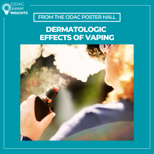
Electronic cigarette use is increasing in the U.S., rising from 4.5% in 2019 to 6.5% in 2023. Although often perceived as safer than traditional smoking, vaping exposes users to carcinogens, volatile compounds, and heavy metals, and its long-term health effects are not fully known. A scoping review presented as a poster at ODAC examines the dermatologic and oral health impacts of vaping.

In an interview with Next Steps in Derm, poster author Julianna Gregory, BSN, shares that the most common findings involved the face and oral cavity. Patients developed persistent oral ulcerations that often improved only after stopping vaping. Facial effects included irritant and allergic contact dermatitis of the lips, perioral skin, cheeks, and eyelids, sometimes linked to metal allergens such as nickel from device components. Vaping was also reported to exacerbate inflammatory and autoimmune skin diseases, including discoid lupus.

Beyond the face, vaping was associated with urticaria and diffuse inflammatory eruptions on the trunk and extremities, suggesting systemic immune activation. Hand dermatitis was also reported due to repeated contact with devices and e-liquid residue. A case of post-surgical free flap compromise highlighted potential vasoconstrictive effects of vaping and raised concerns about impaired wound healing, similar to traditional smoking.

Importantly, dermatologic reactions were reported even with nicotine-free products, indicating that other constituents—such as flavorings, propylene glycol, glycerin, thermal byproducts, and device-derived materials—can act as irritants, allergens, or immune triggers.

Clinicians should routinely ask about vaping as part of the dermatologic history, consider it in unexplained or treatment-resistant conditions (especially involving the face, hands, or oral mucosa), and counsel patients on cessation, including in perioperative settings. The findings also highlight the need for better documentation of vaping behaviors and greater awareness of its potential cutaneous risks.

AD and Regional Eczemas: ODAC in the News

By ODAC Sessions

AD and regional eczemas

At the ODAC Dermatology Conference, Peter Lio, MD, FAAD, shared an updated, patient-centered overview of atopic dermatitis (AD) and regional eczemas, integrating emerging science with real-world clinical strategies.

As noted in an article by The Dermatology Digest, Dr. Lio emphasized AD and regional eczemas are self-perpetuating disorders involving the skin barrier, immune system, microbiome, and sensory nerves. He outlined the epithelial barrier hypothesis and the concept of epicutaneous sensitization. Early, proactive intervention was framed as essential to preventing chronic inflammation, infections, and subsequent disease.

On therapy, Dr. Lio reviewed the expanding role of non-steroidal topical agents, noting their usefulness in sensitive areas and for patients who want a non-steroidal option. He also reinforced the value of adjunctive strategies such as wet wrap therapy. For systemic treatment, he discussed biologics and oral JAK inhibitors, highlighting evidence that dupilumab can uniquely improve the skin microbiome in moderate-to-severe AD. Shared decision-making was a central theme, with Dr. Lio presenting his ESTAR framework to help align treatment choices with patient priorities, and sharing the importance of written action plans.

Finally, Dr. Lio addressed topical steroid withdrawal as a legitimate and evolving diagnosis. He noted that formal diagnostic criteria are forthcoming, signaling progress toward clearer guidance for clinicians.

Evaluating Skin Findings of Systemic Disease

By ODAC Sessions

skin findings of systemic disease

At the 2026 ODAC Dermatology Conference, attendees had the opportunity to learn about evaluating skin findings of systemic disease from Olayemi Sokumbi, MD, FAAD, professor of dermatology and laboratory medicine & pathology at Mayo Clinic. Dr. Sokumbi shared her expertise about evaluating skin findings that may occur in the context of systemic illness, emphasizing a structured derm-dermpath approach. Through two illustrative cases, she demonstrated how the skin serves as an early window to systemic disease and how clinicopathologic correlation (CPC) is integral to establishing the correct diagnosis.

Case 1:

Dr. Sokumbi described a gentleman in his 50s who presented with skin-colored papules on the ears accompanied by joint pains. The presence of concurrent systemic symptoms raised suspicion for an underlying systemic process, prompting a skin biopsy. Histopathology revealed foamy xanthomatous histiocytes, suggesting a non-Langerhans cell histiocytosis with inflammatory arthritis such as Erdheim-Chester disease (EDC). However, at least half of cases of EDC cases demonstrate a BRAF V600E mutation.1 Staining of this skin biopsy was negative for this mutation, prompting Dr. Sokumbi to return to the bedside and broaden the differential diagnosis. Subsequent physical examination revealed periungual papules and nodules, in a characteristic “coral beading” pattern, leading to the diagnosis of multicentric reticulohistiocytosis (MRH), a condition also associated with severe polyarthritis. The histopathologic pitfall requiring CPC was the presence of xanthomatized histiocytes, which are typical for EDC and underrecognized in MRH due to the rarity of this finding. Accurate diagnosis carries significant clinical implications, as MRH has a strong association with solid organ malignancy and requires therapeutic approaches distinct from those used in EDC.

Case 2:

Dr. Sokumbi presented the case of a young lady with diffuse cutaneous hyperpigmentation, which multiple providers had attributed to dermatoheliosis or photoaging. She highlighted, however, key photoprotected areas, such as the conchal bowls of the ears, also demonstrated blue-gray discoloration. The clinical differential diagnoses included lichen planus pigmentosus and argyria, yet the characteristic histopathologic features of these entities were not present on skin biopsy.

Instead, histologic examination revealed wavy deposits within the dermis that stained basophilic on Hematoxylin and Eosin and blue-black with Verhoeff Van Gieson (VVG) staining. Basophilic collagen fibers and altered deposits of elastic fibers have been reported as early-stage findings of ochronosis,3 in contrast to the classic late-stage yellow-brown banana-shaped collagen fibers. Based on these findings the patient was diagnosed with endogenous ochronosis/alkaptounuria, a genodermatosis characterized by impaired breakdown of tyrosine and phenylalanine.

In conclusion, Dr. Sokumbi emphasized how dermatologists are often uniquely positioned to diagnose systemic disease through careful evaluation of skin findings. Both cases underscored the importance of CPC. She encouraged repeating skin biopsies when the leading diagnosis remains unclear and collaborating with colleagues across specialties to ensure comprehensive management of systemic disease.

This session summary was written by Nagasai Adusumilli, MD, MBA, chief resident physician in dermatology at the George Washington University School of Medicine and Health Sciences.

References

  1. Haroche J, Cohen-Aubart F, Emile JF, et al. Reproducible and sustained efficacy of targeted therapy with vemurafenib in patients with BRAF(V600E)-mutated Erdheim-Chester disease. J Clin Oncol. 2015 Feb 10;33(5):411-8. PMID: 25422482.
  2. Camargo K, Pinkston O, Abril A, Sluzevich JC. Xanthomatous multicentric reticulohistiocytosis: an underrecognized variant. J Clin Rheumatol. 2018 Aug;24(5):285-287. PMID: 29239933.
  3. Chowdary S, Mahalingam M, Vashi NA. Reading between the layers: early histopathological findings in exogenous ochronosis. Am J Dermatopathol. 2014 Dec;36(12):989-91.PMID: 25415140.

Pain Management & Wound Care in HS

By ODAC Sessions

pain management in hidradenitis suppurativa

At the 2026 ODAC Dermatology Conference in Orlando, hidradenitis suppurativa (HS) expert Hadar Lev-Tov, MD, gave a high-yield, practice-focused lecture on two frequently underappreciated pillars of HS care: pain management and wound care. According to Dr. Lev-Tov, addressing these areas is essential to improving quality of life and overall outcomes for patients with HS.

Pain as a Core Component of HS Care
Dr. Lev-Tov emphasized that pain is not a secondary symptom of HS but a central driver of disease burden. A large proportion of patients experience moderate to severe pain, which often impacts quality of life more than objective disease severity. Despite this, pain remains underrecognized and undertreated in dermatology clinics. Because dermatologists serve as the primary physicians for most HS patients, Dr. Lev-Tov stressed that pain management must be viewed as an integral part of HS care. Uncontrolled pain also increases the risk of long-term opioid use, an association that persists even after adjusting for confounders.

A Multimodal, Bio-Psycho-Social Approach
To address chronic HS pain, Dr. Lev-Tov advocated for a multimodal strategy rooted in a bio-psycho-social framework. Pharmacologic management should follow a stepwise approach, beginning with topical or systemic NSAIDs and acetaminophen. For neuropathic pain, agents such as gabapentin, pregabalin, TCAs, and SNRIs (including duloxetine) may be effective. Short courses of tramadol or other short-acting opioids can be considered for acute breakthrough pain, with strict limits on quantity. Procedural interventions, such as intralesional triamcinolone or punch deroofing, and referral to pain specialists should be considered for refractory cases.

Equally important are non-pharmacologic therapies, which form the foundation of chronic pain management. These include physical activation strategies (physical therapy, yoga, low-intensity aerobic exercise), behavioral interventions (cognitive behavioral therapy and mindfulness), and adjunctive techniques (massage, acupuncture, and chiropractic care).

Wound Care: Small Details, Big Impact
The second half of the presentation focused on wound care, which Dr. Lev-Tov described as critical to patient comfort and daily functioning. While medical and surgical treatment address disease control, appropriate wound care can dramatically improve quality of life—and poor dressing choices can undermine even the best treatment plans.

Key pearls included selecting dressings based on wound depth and exudate level, with practical guidance on the use of foams, hydrocolloids, gelling fibers, calcium alginates, and superabsorbent dressings. Dr. Lev-Tov also highlighted evidence supporting wide excision with healing by secondary intent, noting excellent outcomes even for large HS wounds. Dedicated HS wound care systems, such as HidraWear, can further enhance comfort, confidence, and ease of use.

Additional surgical pearls included continuing biologic therapy through HS surgery, recognizing and managing hypergranulation tissue, and using chemical debridement, topical corticosteroids, or mechanical debulking when needed.

Takeaway
Dr. Lev-Tov’s lecture on pain management and wound care in HS reinforced that effective HS care extends beyond inflammation control. Pain management is essential—not optional—and thoughtful, individualized wound care plays a major role in improving patient quality of life. By embracing a multimodal pain strategy and a structured approach to wound dressing selection, dermatologists can make a meaningful difference for patients living with HS.

This session summary was written by Dr. Ryan Gall and published on Next Steps in Derm.