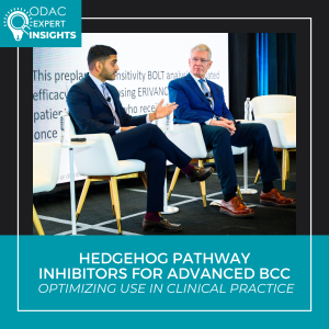
At the 2026 ODAC Conference, an expert panel led by Vishal A. Patel, MD, FACMS, and C. William Hanke, MD, MPH, FAAD, reviewed evolving strategies for hedgehog pathway inhibitors (HHIs) in advanced basal cell carcinoma (BCC), emphasizing practical clinical decision-making.
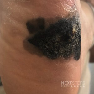
The discussion highlighted the two FDA-approved HHIs: vismodegib (approved in 2012 following the ERIVANCE trial) and sonidegib (approved in 2015 based on the BOLT trial). Reported objective response rates were 47% for vismodegib and 56.1% for sonidegib, with median durations of response of 9.5 months and 26 months, respectively. While cross-trial comparisons are limited, both agents demonstrated meaningful disease control and durable responses with continued therapy.
HHIs offer significant tumor shrinkage and may allow retreatment, but they are not typically curative. Common class-related adverse effects—including muscle cramps, dysgeusia, alopecia, and gastrointestinal symptoms—are usually low grade yet chronic, often affecting quality of life and adherence. Sonidegib’s longer half-life (28–30 days) compared with vismodegib (4–12 days) may influence toxicity duration and management decisions.
Clinically, HHIs are best used selectively:
- As primary therapy for unresectable or inoperable BCC
- As neoadjuvant therapy to reduce tumor burden before surgery or radiation
- As bridge therapy to optimize cosmetic or functional outcomes
Even without complete tumor eradication, many patients achieve meaningful functional and cosmetic improvement. Ultimately, successful use of hedgehog inhibitors for advanced basal cell carcinoma depends on careful patient selection, proactive management of adverse effects, and thoughtful integration with surgery, radiation, and immunotherapy—expanding dermatologists’ options beyond surgical management alone.
This session summary was written by Dr. Erica Lin and published on Next Steps in Derm.