
Category

ODAC Sessions

GLP-1 Agonists in Cutaneous Medicine: Rapid-Fire Insights

By ODAC Sessions

GLP-1 agonists in dermatology

At the ODAC Dermatology Conference, Drs. Jennifer Soung, Steven Daveluy, and Sarah Jackson joined moderator Dr. Adam Friedman for a rapid-fire discussion on GLP-1 receptor agonists in dermatology. “You honestly have to be living under a rock not to see the excitement around GLP-1s,” Dr. Friedman opened, noting their transformative impact across chronic disease management and weight loss.

Psoriasis highlights the link between obesity and systemic inflammation. “Psoriasis is intimately related to obesity,” Dr. Soung emphasized. “The higher the BMI, the higher the PASI score. GLP-1s aren’t just powerful for weight loss—they also improve blood pressure, glycemic control, cardiovascular outcomes.” She added, “Obesity is not a matter of choice; it is a neurohormonal disease.”

Weight loss may also enhance treatment outcomes. “One recent study showed that obesity can reduce the efficacy of biologics,” Soung noted. She highlighted that GLP-1–induced weight loss parallels benefits seen in low-calorie diets but without the extreme patient burden. “Patients can now lose more than 10% of their weight besides bariatric surgery,” she said, underscoring the systemic and potential anti-inflammatory benefits of GLP-1 agonists.

Hidradenitis suppurativa (HS) reflects similar comorbidity patterns. Dr. Daveluy stated, “Obesity is a pro-inflammatory state. The interplay of inflammation and adipocytes can flare chronic skin disease.” He cited early studies showing that GLP-1 agonists improved HS outcomes, including modified Hurley stage and Dermatology Life Quality Index scores, sometimes independent of weight loss. “These agonists activate parts of the immune system and can decrease TNF, IL-17, and IL-23,” Daveluy explained.

The panel also explored aesthetic implications. “Massive weight loss impacts dermal white adipose tissue and facial structure,” said Dr. Jackson. She emphasized early intervention with fillers and biostimulatory treatments: “Start early—inject before patients experience facial volume loss. Target not only the skin but adipocytes and volume.” She also stressed patient counseling: “Set expectations—this isn’t a one-tube-of-filler journey. Go low and slow on GLP-1 doses, and support protein intake for hair and muscle.”

Hair loss emerged as a frequently reported adverse effect. “We see initial telogen effluvium and, surprisingly, early androgenic-like thinning,” noted Dr. Friedman. He recommended proactive measures: “Start oral or topical minoxidil early. Anti-androgen therapy can be considered, but increased blood flow through minoxidil is a great first step.”

Dr. Friedman concluded by summarizing one key takeaway: “Obesity and adipocyte dysfunction create a pro-inflammatory state that worsens chronic dermatologic disease. Removing that burden can have a positive impact, but everything has a cost.” The panel emphasized vigilance, early intervention, and coordinated care to maximize benefits while mitigating adverse effects such as lipoatrophy or hair thinning.

This information was presented at the 2026 ODAC conference by Steven Daveluy, MD, FAAD; Adam Friedman, MD, FAAD; Sarah Jackson, MD, FAAD; and Jennifer Soung, MD, FAAD. The above highlights from this lecture were written and compiled by Samip Sheth, MD.

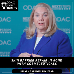
Skin Barrier Repair in Acne with Cosmeceuticals

By ODAC Sessions

skin barrier repair in acne

Acne is a barrier deficiency disorder, according to Brooklyn, N.Y., dermatologist Dr. Hilary Baldwin. Next Steps in Derm, in partnership with the ODAC Dermatology Conference, interviewed Dr. Baldwin, who addresses the role of skin care in acne improvement. Find out why better products and vehicles could be the reason for the increasing placebo effect in acne studies. Find out the impact of acne treatments on the skin barrier and how this affects non-compliance. Hear what Dr. Baldwin says must happen in every acne visit. Plus learn the origin of the new term “acneceuticals” and what the latest research shows on the role of non-prescription active ingredients in acne care.


Further Reading

If you want to read more about cosmeceuticals for acne, check out the following articles published in the Journal of Drugs in Dermatology:

The Use of Acneceuticals to Improve Acne Care: Introduction of a New Term and Review of the Literature

ABSTRACT

Background: Acne is a multifactorial inflammatory skin condition that commonly presents to the dermatology clinic. Treatment generally involves the use of pharmaceutical agents and procedural techniques. Recently, the importance of over-the-counter skin care in acne

has been recognized in many studies. This paper introduces the term acneceuticals to encompass a wide range of FDA monographed, yet non-prescription ingredients proven to alter the structure and function of acneic skin.

Methods: A panel of 8 dermatologists with an interest in acne and skin care performed a literature review of active skin care in acne. The role of acneceuticals in the treatment of acne — as monotherapy, adjunctive, and maintenance therapy — was evaluated using a modified Delphi approach. Studies were limited to in vivo human trials involving acne. Individual actives were assessed separately.

Results: The quality of evidence was moderate-to-low for many of the ingredients. Most of the actives included in the final assessment had been studied in vehicle-controlled, blinded, often comparative studies but enrolled a small number of subjects. In these studies, the acneceuticals were found to reduce lesion count, reduce sebum production, and improve efficacy of existing pharmacologic therapies.

Conclusion: Acneceuticals have demonstrated benefits in treating acne, alone or in conjunction with established pharmaceutical agents. These data allow us to make quality recommendations for our patients that should be a part of every patient encounter. The recommendations also serve as a guide for patients searching the internet for beneficial self-care products.

Real-World Clinical Case Series Utilizing Acneceuticals as Monotherapy, Adjunctive, or Maintenance Therapy for Acne Vulgaris

ABSTRACT

Background: Acne vulgaris is a common, multifactorial inflammatory skin disease for which there are many pharmacologic and procedural interventions. Recent publications have stressed the importance of quality skin care containing non-prescription actives (acneceuticals) in the treatment of this chronic disorder. Acne therapy is made more complicated by the diversity of presentation that includes age, gender, race, underlying skin type, product adherence, lesion morphology, and severity.

Methods: A panel of 6 dermatologists with expertise in the treatment of acne met twice for consensus conferences in March and November 2024. Discussion included the generation of 13 common disease presentations, the identification of potential cases to illustrate the presentations, and the design of 2-month case studies utilizing acneceuticals. Panelists chose new or established patients in their care who fit into the predetermined categories, provided them with the agreed-upon acneceuticals, and followed them for 2 months.

Results: Ten cases were chosen to comprise the most common therapeutic crossroads where pharmacologic or procedural interventions were not providing satisfactory control. This case study series included patients who were medication-intolerant, failing internet cures, not fully responsive to their current regimen, adult female acne patients with concomitant aging concerns, those needing maintenance therapy, and patients with issues regarding access to care.

Conclusions: Acneceuticals were found to play an important role as monotherapy, adjunctive therapy, and maintenance therapy for acne patients. The 10 cases elucidated the ability of these over-the-counter (OTC) actives to improve patient care under several clinical scenarios.

Did you enjoy this video interview? Find more here.
Platelet-Rich Plasma and Procedural Interventions for Androgenetic Alopecia

By ODAC Sessions

procedural interventions for androgenetic alopecia

At the ODAC Dermatology Conference, Terrence C. Keaney, MD, delivered a practical, experience-driven update on procedural interventions for androgenetic alopecia (AGA), with a primary focus on platelet-rich plasma (PRP).

Dr. Keaney opened by reframing treatment escalation. “Hair loss is one of the few conditions where the severity of disease does not dictate how aggressively you treat,” he said. “It depends on how much the patient is bothered.” In his practice, patients rate distress from 0 to 10 at intake.

While emphasizing that medical therapy remains foundational, he described PRP as the “mainstay regenerative option” in his clinic. Mechanistically, PRP concentrates platelets that release a “cocktail of growth factors” and cytokines. “If we could isolate the single magic molecule, we’d have a drug,” he noted. “But right now, it’s the combined signal that appears to prevent dermal papilla apoptosis and prolong anagen.”

He acknowledged that data show a treatment signal but stressed variability. “Not everyone responds, and you have to be straightforward about that.” One major factor is biologic fluctuation. “The ‘drug’ in PRP is what’s in the alpha granules of platelets,” he said. “But platelet counts vary by time of day, hydration, diet, age, and sex.” A fasting, highly hydrated morning patient may yield a lower effective concentration than an afternoon, well-fed patient. “You may be delivering different doses without realizing it.”

Preparation differences add further inconsistency. “Not one PRP study uses the exact same protocol,” he said, citing variability in centrifugation, leukocyte content, activation, concentration targets, and treatment intervals. While many clinicians aim for 5–10× physiologic platelet concentration, he emphasized that no single protocol is definitively superior.

In his practice, Dr. Keaney performs three monthly “loading” treatments followed by maintenance every three to six months. “That’s based on experience, not perfect science,” he acknowledged.

On safety, he referenced reports of vision loss after periocular PRP injections. Although no cases have been reported with scalp injections, he remains cautious. “You’re injecting a concentrated clot,” he said, underscoring the importance of vascular awareness and informed consent.

He briefly addressed alternatives. Platelet-rich fibrin (PRF), he suggested, may be less biologically rational for hair loss. On exosomes, he noted the U.S. Food and Drug Administration considers injectable exosomes a drug. “Do not inject exosomes,” he cautioned.

Finally, Dr. Keaney emphasized objective tracking. His clinic uses standardized photography and trichoscopic measurements. “The follow-up becomes easy,” he said. “You can show the patient real data—no debating lighting or styling.”

His takeaway for dermatologists: Procedural therapies can augment medical management of AGA, but success depends on careful patient selection, transparency about variability, and disciplined technique.

This summary was prepared by Dr. Samip Sheth, dermatology resident, who attended the session. The content reflects the resident’s notes and interpretations, may contain errors, and is provided for educational purposes only. It does not constitute official faculty endorsement and should not replace original sources or clinical judgment.


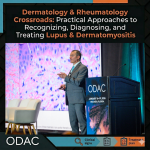
Recognizing and Treating Cutaneous Lupus and Dermatomyositis

By ODAC Sessions

autoimmune connective tissue disorders

At the ODAC Dermatology Conference, Anthony Fernandez, MD, PhD, FAAD, shared his experience in autoimmune connective tissue disorders, highlighting the intersection between dermatology and rheumatology, and underscoring the importance of practical approaches in complex diseases.

Cutaneous Lupus

Dr. Fernandez began his session by addressing cutaneous lupus, stressing the need to recognize lesion patterns. “If you become familiar with the morphology of the skin lesions and the pattern of distribution, you should be able to recognize lupus and dermatomyositis.”

Regarding acute cutaneous lupus, he noted that, “All of these patients have systemic lupus erythematosus (SLE), so [they] are going to have a positive ANA and usually numerous positive anti-ENA antibodies.” However, he also cautioned, “Not every patient has a classic butterfly rash, so you need to look at the entire clinical picture.”

For subacute cutaneous lupus (SCLE), he emphasized photosensitivity. “Annular scaly lesions or psoriasiform lesions in a photo-distribution are a hallmark. About 25-40% of these patients may develop SCLE because of a medication they’re taking, so you need to perform a good medication history upon diagnosis.”

Chronic cutaneous lupus, particularly discoid lupus (DLE), carries unique implications. “A negative ANA does not rule out cutaneous lupus, especially with discoid lesions,” he stressed. “Patients with generalized discoid lupus, meaning they have lesions both above and below the neck, are at increased risk for progressing to SLE compared to patients with DLE lesions only above the neck.”

Dr. Fernandez advised antimalarials, hydroxychloroquine, and chloroquine as first-line systemic treatments for cutaneous lupus. “If a patient fails the first antimalarial, there is research supporting that switching to the alternative antimalarial may be effective in a significant percentage of cases.”

Dermatomyositis

Turning to dermatomyositis, Dr. Fernandez highlighted hallmark cutaneous features: heliotrope rash, Gottron’s papules, shawl sign, V-neck sign, and holster sign. He noted that any combination of these features may occur in patients with dermatomyositis, and cautioned about MDA5-positive disease. “These patients often present with skin lesions due to vasculopathy. There is a very strong risk of interstitial lung disease (ILD). Some may have a rapidly progressive ILD phenotype, which can be life-threatening despite aggressive treatment.”

For diagnosis, he recommended combining clinical and objective data. “You need to correlate what you see clinically with some objective data, mainly a lesional skin biopsy with histopathologic characteristics consistent with lupus or dermatomyositis.” He also advised autoantibody testing. “For dermatomyositis, myositis-specific autoantibodies correlate with clinical phenotype, so they have tremendous value in terms of how you test, monitor, and treat these patients.”

For treatment, Dr. Fernandez recommended typically starting with corticosteroids plus a steroid-sparing agent. “If patients fail traditional agents, IVIG is far and away the best medicine we currently have for treating the skin, myositis, or both.”

Looking ahead, he concluded with excitement about emerging therapies. “Oral JAK inhibitors, anti-interferon receptor monoclonal antibodies, and anti-interferon-beta monoclonal antibodies may be the first truly novel medicines that get approved for dermatomyositis in decades, depending upon results of ongoing phase 3 trials.” He conveyed to attendees, “Once you make a diagnosis, we currently have good medicines to offer, but the real excitement is what’s in the pipeline right now.”

This information was presented at the 2026 ODAC conference by Anthony Fernandez, MD, PhD, FAAD. The above highlights from this lecture were written and compiled by Samip Sheth, MD.

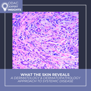
A Dermatology & Dermatopathology Approach to Systemic Disease

By ODAC Sessions

clinicopathologic correlation

At ODAC, Olayemi Sokumbi, MD, FAAD, delivered a masterclass in clinicopathologic correlation — a timely reminder that careful clinicopathologic thinking changes diagnoses, management, and outcomes. Through two cases, Dr. Sokumbi outlined how clinicopathologic correlation is a clinical imperative:

  • Case 1: Flesh‑colored papules initially called “skin tags” were reclassified as multicentric reticulohistiocytosis after integrating distribution, clinical course (periungual/hand involvement, inflammatory arthritis), and histology — a diagnosis with major implications for arthritis management and malignancy screening.
  • Case 2: Subtle, diffuse skin discoloration with otherwise non‑diagnostic biopsies was clarified by elastic tissue stain to be ochronosis due to alkaptonuria, prompting genetic and systemic workup.

These cases illustrate how dermatologists and dermatopathologists can be the first to uncover multisystem disease and steer timely, life‑altering care.

Practical reminders: Don’t be afraid to re‑biopsy, use targeted IHC and special stains, and maintain close dermatopathology collaboration. Persistence and multidisciplinary care are often the key.

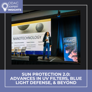
Advances in UV Filters, Blue Light Defense, & Beyond

By ODAC Sessions

sun protection

At ODAC, Misty Eleryan, MD, MS, FACMS, delivered a timely roadmap for Sun Protection 2.0 — reframing photoprotection beyond UV and into visible/infrared damage, formulation science, and personalized strategies.

Key takeaways:

  • Photodamage spans UVA/UVB, visible light, and infrared — explaining why some patients still photoage despite diligent sunscreen use.
  • White cast from mineral sunscreens undermines adherence; improving texture and cosmetic elegance is a public-health priority.
  • Innovation is underway: nanotechnology for photostability and reduced absorption, antioxidants (vitamins C/E) to neutralize reactive oxygen species, sunscreen “boosters,” and potential biotech discovery of novel UV filters.
  • Regulatory reality: no new US filters since 1999. The 2025 SAFE Sunscreen Standards Act may finally accelerate safe, globally informed approvals.
  • Emerging tools: wearable UV sensors, topical cannabidiol research, and oral photoprotective supplements like polypodium.

Practical message: Sunscreen remains the foundation, but photoprotection must be multifaceted, involving product choice, behavior, nutrition, and emerging tech. Dermatologists should lead these conversations to improve adherence and outcomes.

Read the full session summary written by Milaan Shah, MD, to explore formulation advances, policy shifts, and actionable counseling tips you can use with patients today.

Rethinking Prurigo Nodularis: ODAC Session Summary

By ODAC Sessions

prurigo nodularis

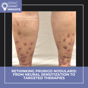
At ODAC 2026, Adam Friedman, MD, FAAD, shared a prurigo nodularis (PN) case that might feel familiar: multiple nodules, relentless itch, one question — why? His message: PN is not merely scratching or a bystander to other dermatoses. It’s a distinct neuroimmune disorder with systemic implications, and overlap does not mean sameness.

PN is sustained by a maladaptive neuroimmune loop: IL‑31 drives itch via sensory neurons, periostin amplifies neural signaling and keratinocyte inflammation, and chronic scratching remodels skin and nerves. Clinically this maps to two meaningful subtypes — inflammatory‑predominant and neuropathic‑predominant — which helps explain variable responses to therapy.

Management goals are simple: reduce itch, interrupt the itch‑scratch cycle, and heal lesions. Tools span topical agents (emerging data for ruxolitinib), neural modulators (gabapentin, pregabalin, mirtazapine), adjuncts like PEA, targeted biologics (nemolizumab), and evolving JAK inhibitors. Expectation setting is key: control, not cure, and “stay on to stay clear” for many patients.

PN often coexists with systemic disease and lifestyle factors that compound burden and reduce quality of life. As mechanism‑driven therapies expand, identifying phenotype and personalizing combinations will improve outcomes.

Read the full session summary, written by Tammy Gonzalez, MD, PhD, to dive into Dr. Friedman’s practical framework and therapeutic insights.

AD and Regional Eczemas: ODAC in the News

By ODAC Sessions

AD and regional eczemas

At the ODAC Dermatology Conference, Peter Lio, MD, FAAD, shared an updated, patient-centered overview of atopic dermatitis (AD) and regional eczemas, integrating emerging science with real-world clinical strategies.

As noted in an article by The Dermatology Digest, Dr. Lio emphasized AD and regional eczemas are self-perpetuating disorders involving the skin barrier, immune system, microbiome, and sensory nerves. He outlined the epithelial barrier hypothesis and the concept of epicutaneous sensitization. Early, proactive intervention was framed as essential to preventing chronic inflammation, infections, and subsequent disease.

On therapy, Dr. Lio reviewed the expanding role of non-steroidal topical agents, noting their usefulness in sensitive areas and for patients who want a non-steroidal option. He also reinforced the value of adjunctive strategies such as wet wrap therapy. For systemic treatment, he discussed biologics and oral JAK inhibitors, highlighting evidence that dupilumab can uniquely improve the skin microbiome in moderate-to-severe AD. Shared decision-making was a central theme, with Dr. Lio presenting his ESTAR framework to help align treatment choices with patient priorities, and sharing the importance of written action plans.

Finally, Dr. Lio addressed topical steroid withdrawal as a legitimate and evolving diagnosis. He noted that formal diagnostic criteria are forthcoming, signaling progress toward clearer guidance for clinicians.

Evaluating Skin Findings of Systemic Disease

By ODAC Sessions

skin findings of systemic disease

At the 2026 ODAC Dermatology Conference, attendees had the opportunity to learn about evaluating skin findings of systemic disease from Olayemi Sokumbi, MD, FAAD, professor of dermatology and laboratory medicine & pathology at Mayo Clinic. Dr. Sokumbi shared her expertise about evaluating skin findings that may occur in the context of systemic illness, emphasizing a structured derm-dermpath approach. Through two illustrative cases, she demonstrated how the skin serves as an early window to systemic disease and how clinicopathologic correlation (CPC) is integral to establishing the correct diagnosis.

Case 1:

Dr. Sokumbi described a gentleman in his 50s who presented with skin-colored papules on the ears accompanied by joint pains. The presence of concurrent systemic symptoms raised suspicion for an underlying systemic process, prompting a skin biopsy. Histopathology revealed foamy xanthomatous histiocytes, suggesting a non-Langerhans cell histiocytosis with inflammatory arthritis such as Erdheim-Chester disease (EDC). However, at least half of cases of EDC cases demonstrate a BRAF V600E mutation.1 Staining of this skin biopsy was negative for this mutation, prompting Dr. Sokumbi to return to the bedside and broaden the differential diagnosis. Subsequent physical examination revealed periungual papules and nodules, in a characteristic “coral beading” pattern, leading to the diagnosis of multicentric reticulohistiocytosis (MRH), a condition also associated with severe polyarthritis. The histopathologic pitfall requiring CPC was the presence of xanthomatized histiocytes, which are typical for EDC and underrecognized in MRH due to the rarity of this finding. Accurate diagnosis carries significant clinical implications, as MRH has a strong association with solid organ malignancy and requires therapeutic approaches distinct from those used in EDC.

Case 2:

Dr. Sokumbi presented the case of a young lady with diffuse cutaneous hyperpigmentation, which multiple providers had attributed to dermatoheliosis or photoaging. She highlighted, however, key photoprotected areas, such as the conchal bowls of the ears, also demonstrated blue-gray discoloration. The clinical differential diagnoses included lichen planus pigmentosus and argyria, yet the characteristic histopathologic features of these entities were not present on skin biopsy.

Instead, histologic examination revealed wavy deposits within the dermis that stained basophilic on Hematoxylin and Eosin and blue-black with Verhoeff Van Gieson (VVG) staining. Basophilic collagen fibers and altered deposits of elastic fibers have been reported as early-stage findings of ochronosis,3 in contrast to the classic late-stage yellow-brown banana-shaped collagen fibers. Based on these findings the patient was diagnosed with endogenous ochronosis/alkaptounuria, a genodermatosis characterized by impaired breakdown of tyrosine and phenylalanine.

In conclusion, Dr. Sokumbi emphasized how dermatologists are often uniquely positioned to diagnose systemic disease through careful evaluation of skin findings. Both cases underscored the importance of CPC. She encouraged repeating skin biopsies when the leading diagnosis remains unclear and collaborating with colleagues across specialties to ensure comprehensive management of systemic disease.

This session summary was written by Nagasai Adusumilli, MD, MBA, chief resident physician in dermatology at the George Washington University School of Medicine and Health Sciences.

References

  1. Haroche J, Cohen-Aubart F, Emile JF, et al. Reproducible and sustained efficacy of targeted therapy with vemurafenib in patients with BRAF(V600E)-mutated Erdheim-Chester disease. J Clin Oncol. 2015 Feb 10;33(5):411-8. PMID: 25422482.
  2. Camargo K, Pinkston O, Abril A, Sluzevich JC. Xanthomatous multicentric reticulohistiocytosis: an underrecognized variant. J Clin Rheumatol. 2018 Aug;24(5):285-287. PMID: 29239933.
  3. Chowdary S, Mahalingam M, Vashi NA. Reading between the layers: early histopathological findings in exogenous ochronosis. Am J Dermatopathol. 2014 Dec;36(12):989-91.PMID: 25415140.

Pain Management & Wound Care in HS

By ODAC Sessions

pain management in hidradenitis suppurativa

At the 2026 ODAC Dermatology Conference in Orlando, hidradenitis suppurativa (HS) expert Hadar Lev-Tov, MD, gave a high-yield, practice-focused lecture on two frequently underappreciated pillars of HS care: pain management and wound care. According to Dr. Lev-Tov, addressing these areas is essential to improving quality of life and overall outcomes for patients with HS.

Pain as a Core Component of HS Care
Dr. Lev-Tov emphasized that pain is not a secondary symptom of HS but a central driver of disease burden. A large proportion of patients experience moderate to severe pain, which often impacts quality of life more than objective disease severity. Despite this, pain remains underrecognized and undertreated in dermatology clinics. Because dermatologists serve as the primary physicians for most HS patients, Dr. Lev-Tov stressed that pain management must be viewed as an integral part of HS care. Uncontrolled pain also increases the risk of long-term opioid use, an association that persists even after adjusting for confounders.

A Multimodal, Bio-Psycho-Social Approach
To address chronic HS pain, Dr. Lev-Tov advocated for a multimodal strategy rooted in a bio-psycho-social framework. Pharmacologic management should follow a stepwise approach, beginning with topical or systemic NSAIDs and acetaminophen. For neuropathic pain, agents such as gabapentin, pregabalin, TCAs, and SNRIs (including duloxetine) may be effective. Short courses of tramadol or other short-acting opioids can be considered for acute breakthrough pain, with strict limits on quantity. Procedural interventions, such as intralesional triamcinolone or punch deroofing, and referral to pain specialists should be considered for refractory cases.

Equally important are non-pharmacologic therapies, which form the foundation of chronic pain management. These include physical activation strategies (physical therapy, yoga, low-intensity aerobic exercise), behavioral interventions (cognitive behavioral therapy and mindfulness), and adjunctive techniques (massage, acupuncture, and chiropractic care).

Wound Care: Small Details, Big Impact
The second half of the presentation focused on wound care, which Dr. Lev-Tov described as critical to patient comfort and daily functioning. While medical and surgical treatment address disease control, appropriate wound care can dramatically improve quality of life—and poor dressing choices can undermine even the best treatment plans.

Key pearls included selecting dressings based on wound depth and exudate level, with practical guidance on the use of foams, hydrocolloids, gelling fibers, calcium alginates, and superabsorbent dressings. Dr. Lev-Tov also highlighted evidence supporting wide excision with healing by secondary intent, noting excellent outcomes even for large HS wounds. Dedicated HS wound care systems, such as HidraWear, can further enhance comfort, confidence, and ease of use.

Additional surgical pearls included continuing biologic therapy through HS surgery, recognizing and managing hypergranulation tissue, and using chemical debridement, topical corticosteroids, or mechanical debulking when needed.

Takeaway
Dr. Lev-Tov’s lecture on pain management and wound care in HS reinforced that effective HS care extends beyond inflammation control. Pain management is essential—not optional—and thoughtful, individualized wound care plays a major role in improving patient quality of life. By embracing a multimodal pain strategy and a structured approach to wound dressing selection, dermatologists can make a meaningful difference for patients living with HS.

This session summary was written by Dr. Ryan Gall and published on Next Steps in Derm.