All Posts By

Allison Sit

Launching a Dermatology Practice: ODAC in the News

By Media Coverage

starting a dermatology practice

For many dermatologists, transitioning from an academic medical center or an employed position to owning a private practice is the ultimate career milestone. Yet trading a clinical template for an entrepreneurial business plan can feel like entering uncharted territory.

At the 2026 ODAC Dermatology Conference, Ronda Farah, MD, FAAD, associate professor at the University of Minnesota and founder of Alluma Dermatology, shared a roadmap for clinicians looking to make the leap.

In a recent The Dermatology Digest article, Dr. Farah outlines the foundational pillars required to successfully build a dermatology practice from the ground up. From developing a business plan and securing financing to reviewing your current employment contract, Dr. Farah tells future practice owners what steps to take and what consultants to contact. With an interest in marketing and branding, Dr. Farah also details effective strategies to promote a practice and build a referral network.

Atopic Dermatitis and Mental Health: Patient Buzz

By Medical Dermatology

atopic dermatitis and mental health

Dermatologists are experts at treating what is visible. But as highlighted in a recent article by Everyday Health, there is an “invisible burden” of atopic dermatitis (AD) that cannot be seen on the skin’s surface: the profound toll it takes on a patient’s mental health.

Because AD is a psychophysiological disorder, psychological stress actually triggers inflammation that worsens flares, creating a frustrating, vicious cycle. Chronic itch and sleep disturbances quickly compound this distress, frequently leading to anxiety, depression, and irritability.

Yet a major gap remains in patient care. Patients often hesitate to bring up their mental health during an office visit, and many dermatologists feel unequipped or too pressed for time to broach the topic.

To bridge this gap, read this Next Steps in Derm interview with Mohammad Jafferany, MD, DFAPA, MCPS, a professor of psychodermatology, psychiatry, and behavioral sciences. He shares practical, clinical insights that every dermatologist can implement today. From understanding the bidirectional connection to normalizing the mental health.

GLP-1 Agonists & Psoriasis Comorbidities

By Video Pearls

GLP-1 agonists

Psoriasis is more than skin deep—it is a systemic inflammatory condition. Jennifer Soung, MD, FAAD, argues that addressing metabolic health is no longer outside the scope of dermatology; it is essential for clinical success.

In a Next Steps in Derm video interview at our sister conference Skin of Color Update, Dr. Soung dives into:

  • The Weight-Biologic Link: How excess weight directly impacts the efficacy of biologic therapies.
  • GLP-1 Agonists: Incorporating modern weight management tools into the psoriatic care plan.
  • The Patient Conversation: Strategies for broaching the sensitive topic of weight with empathy and clinical precision.
  • Comorbidity Management: A comprehensive look at tackling psoriatic arthritis and metabolic syndrome.

Stop treating the skin in a vacuum. Learn how to optimize your patients’ systemic health for better dermatologic outcomes.

Recognizing and Treating Cutaneous Lupus and Dermatomyositis

By ODAC Sessions

autoimmune connective tissue disorders

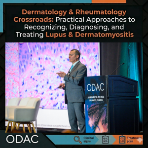
At the ODAC Dermatology Conference, Anthony Fernandez, MD, PhD, FAAD, shared his experience in autoimmune connective tissue disorders, highlighting the intersection between dermatology and rheumatology, and underscoring the importance of practical approaches in complex diseases.

Cutaneous Lupus

Dr. Fernandez began his session by addressing cutaneous lupus, stressing the need to recognize lesion patterns. “If you become familiar with the morphology of the skin lesions and the pattern of distribution, you should be able to recognize lupus and dermatomyositis.”

Regarding acute cutaneous lupus, he noted that, “All of these patients have systemic lupus erythematosus (SLE), so [they] are going to have a positive ANA and usually numerous positive anti-ENA antibodies.” However, he also cautioned, “Not every patient has a classic butterfly rash, so you need to look at the entire clinical picture.”

For subacute cutaneous lupus (SCLE), he emphasized photosensitivity. “Annular scaly lesions or psoriasiform lesions in a photo-distribution are a hallmark. About 25-40% of these patients may develop SCLE because of a medication they’re taking, so you need to perform a good medication history upon diagnosis.”

Chronic cutaneous lupus, particularly discoid lupus (DLE), carries unique implications. “A negative ANA does not rule out cutaneous lupus, especially with discoid lesions,” he stressed. “Patients with generalized discoid lupus, meaning they have lesions both above and below the neck, are at increased risk for progressing to SLE compared to patients with DLE lesions only above the neck.”

Dr. Fernandez advised antimalarials, hydroxychloroquine, and chloroquine as first-line systemic treatments for cutaneous lupus. “If a patient fails the first antimalarial, there is research supporting that switching to the alternative antimalarial may be effective in a significant percentage of cases.”

Dermatomyositis

Turning to dermatomyositis, Dr. Fernandez highlighted hallmark cutaneous features: heliotrope rash, Gottron’s papules, shawl sign, V-neck sign, and holster sign. He noted that any combination of these features may occur in patients with dermatomyositis, and cautioned about MDA5-positive disease. “These patients often present with skin lesions due to vasculopathy. There is a very strong risk of interstitial lung disease (ILD). Some may have a rapidly progressive ILD phenotype, which can be life-threatening despite aggressive treatment.”

For diagnosis, he recommended combining clinical and objective data. “You need to correlate what you see clinically with some objective data, mainly a lesional skin biopsy with histopathologic characteristics consistent with lupus or dermatomyositis.” He also advised autoantibody testing. “For dermatomyositis, myositis-specific autoantibodies correlate with clinical phenotype, so they have tremendous value in terms of how you test, monitor, and treat these patients.”

For treatment, Dr. Fernandez recommended typically starting with corticosteroids plus a steroid-sparing agent. “If patients fail traditional agents, IVIG is far and away the best medicine we currently have for treating the skin, myositis, or both.”

Looking ahead, he concluded with excitement about emerging therapies. “Oral JAK inhibitors, anti-interferon receptor monoclonal antibodies, and anti-interferon-beta monoclonal antibodies may be the first truly novel medicines that get approved for dermatomyositis in decades, depending upon results of ongoing phase 3 trials.” He conveyed to attendees, “Once you make a diagnosis, we currently have good medicines to offer, but the real excitement is what’s in the pipeline right now.”

This information was presented at the 2026 ODAC conference by Anthony Fernandez, MD, PhD, FAAD. The above highlights from this lecture were written and compiled by Samip Sheth, MD.

Acral Dermoscopy: ODAC in the News

By Media Coverage, Uncategorized

acral dermoscopy

The challenge of diagnosing acral lesions—those found on the palms and soles—is a familiar one for even the most seasoned dermatologists. Because these areas follow a unique set of “rules” compared to the rest of the body, clinical confidence is paramount to avoiding unnecessary biopsies while catching early-stage melanomas.

The Dermatology Digest highlights an ODAC session on the topic presented by Jennifer Stein, MD, PhD, FAAD, professor and associate vice chair at the NYU Grossman School of Medicine. The article outlines Dr. Stein’s actionable, everyday strategies, including:

  • The Golden Rule: “Furrows are Fine, Ridges are Risky”
  • Acquired Acral Three-Step Algorithm
  • The BRAAFF Algorithm

In the article, Dr. Stein also shares some considerations in patients with darker skin types:

  • Most algorithms may not perfectly translate to patients with darker skin tones.
  • In Fitzpatrick Skin Types V and VI, clinicians may encounter physiologic volar melanocytic macules that display parallel ridge patterns but are entirely benign. While these findings are common in skin of color, Dr. Stein cautioned that the “index of suspicion” must remain high to ensure acral melanoma isn’t overlooked.

Evaluating the nail unit presents its own set of hurdles, as the pigment often originates in the difficult-to-biopsy nail matrix. Dr. Stein breaks down the evaluation into two categories:

  • Non-melanocytic: Before suspecting a tumor, rule out fungal infections (look for a “reverse triangle” pattern) and subungual hemorrhages (look for red-to-black spots with filamentous edges).
  • Melanocytic: Benign ethnic melanonychia often presents with gray, homogeneous lines. However, the “Ugly Duckling” concept is your best friend here. Look for outliers: bands that are wider at the base, involve multiple colors, or cause nail plate destruction.

Dermoscopy is an invaluable tool, but it works best when paired with clinical intuition. By mastering the distinction between furrows and ridges and remaining mindful of how these patterns shift across different skin types, clinicians can provide more accurate, confident care for their patients.

Read the original article posted to The Dermatology Digest.

Psoriasis and Psoriatic Arthritis in Patients With Skin of Color

By Video Pearls

psoriasis and psoriatic arthritis in patients with skin of color

In a video interview at our sister conference, Skin of Color Update, Mona Shahriari, MD, FAAD, encouraged dermatology clinicians to broaden the psoriasis color palette. She says recognizing the skin tone differences in the presentation of psoriasis can improve diagnosis and care. Dr. Shahriari is associate clinical professor of dermatology at Yale University School of Medicine.

Watch as she shares:

  • The role of biopsy when diagnosing psoriasis in skin of color
  • Challenges in diagnosing psoriatic arthritis in patients with darker skin tones
  • Latest research in treating scalp psoriasis
  • Pigmentary sequelae and the quality of life impact

If you’re committed to providing quality psoriasis care for all of your patients, this is a must-view video.

Topical Nonsteroidal Therapies for Atopic Dermatitis

By Patient Care

topical nonsteroidal therapies

A new consensus in the Journal of Drugs in Dermatology recommends advanced nonsteroidal topicals — topical JAK inhibitors, aryl hydrocarbon receptor agonists, and PDE‑4 inhibitors — as frontline therapy for AD over traditional topical corticosteroids.

In an interview with Next Steps in Derm, author Christopher Bunick, MD, PhD, shares the panel’s evidence‑based process, graded recommendations, and practical implications for clinic.

Why this matters:

  • These agents offer improved efficacy and safety vs. steroids, especially on thin or sensitive skin (face, folds, genitalia).
  • They better control itch, inflammation, and barrier dysfunction — translating to real gains in sleep and quality of life.
  • Their safety profile supports longer‑term use and easier patient adherence.
  • The consensus provides actionable guidance on where nonsteroidal topicals fit alongside biologics and small molecules.

This interview is a must‑read to update your treatment algorithms and patient counseling.

A Dermatology & Dermatopathology Approach to Systemic Disease

By ODAC Sessions

clinicopathologic correlation

At ODAC, Olayemi Sokumbi, MD, FAAD, delivered a masterclass in clinicopathologic correlation — a timely reminder that careful clinicopathologic thinking changes diagnoses, management, and outcomes. Through two cases, Dr. Sokumbi outlined how clinicopathologic correlation is a clinical imperative:

  • Case 1: Flesh‑colored papules initially called “skin tags” were reclassified as multicentric reticulohistiocytosis after integrating distribution, clinical course (periungual/hand involvement, inflammatory arthritis), and histology — a diagnosis with major implications for arthritis management and malignancy screening.
  • Case 2: Subtle, diffuse skin discoloration with otherwise non‑diagnostic biopsies was clarified by elastic tissue stain to be ochronosis due to alkaptonuria, prompting genetic and systemic workup.

These cases illustrate how dermatologists and dermatopathologists can be the first to uncover multisystem disease and steer timely, life‑altering care.

Practical reminders: Don’t be afraid to re‑biopsy, use targeted IHC and special stains, and maintain close dermatopathology collaboration. Persistence and multidisciplinary care are often the key.

Skin Rejuvenation With Microtoxin: From the ODAC Poster Hall

By Aesthetic Dermatology

microtoxin

Microtoxin is shifting how we approach skin rejuvenation — not by paralyzing muscle, but by improving the skin itself. At ODAC, a retrospective chart review presented by Carolyn Duong, BS (Kansas City University), found that superficially placed, highly diluted botulinum toxin produced meaningful gains in texture, pore size, sebum control, reduced erythema, and refinement of shallow scars — all while preserving natural facial movement.

How does it differ from traditional neuromodulators? Microtoxin is delivered as many small intradermal injections across the face to influence sebaceous/eccrine glands, arrector pili, fibroblast signaling, and neurovascular pathways rather than targeting skeletal muscle. The result is diffuse skin quality improvement rather than focal muscle weakening.

Clinical takeaway: Microtoxin is a valuable complementary tool for diffuse skin concerns (texture, oiliness, mild erythema, superficial scars) but is not a replacement for fillers, lasers, or surgery for deep wrinkles, volume loss, or advanced laxity. Technique standardization and careful patient selection are essential.

Advances in UV Filters, Blue Light Defense, & Beyond

By ODAC Sessions

sun protection

At ODAC, Misty Eleryan, MD, MS, FACMS, delivered a timely roadmap for Sun Protection 2.0 — reframing photoprotection beyond UV and into visible/infrared damage, formulation science, and personalized strategies.

Key takeaways:

  • Photodamage spans UVA/UVB, visible light, and infrared — explaining why some patients still photoage despite diligent sunscreen use.
  • White cast from mineral sunscreens undermines adherence; improving texture and cosmetic elegance is a public-health priority.
  • Innovation is underway: nanotechnology for photostability and reduced absorption, antioxidants (vitamins C/E) to neutralize reactive oxygen species, sunscreen “boosters,” and potential biotech discovery of novel UV filters.
  • Regulatory reality: no new US filters since 1999. The 2025 SAFE Sunscreen Standards Act may finally accelerate safe, globally informed approvals.
  • Emerging tools: wearable UV sensors, topical cannabidiol research, and oral photoprotective supplements like polypodium.

Practical message: Sunscreen remains the foundation, but photoprotection must be multifaceted, involving product choice, behavior, nutrition, and emerging tech. Dermatologists should lead these conversations to improve adherence and outcomes.

Read the full session summary written by Milaan Shah, MD, to explore formulation advances, policy shifts, and actionable counseling tips you can use with patients today.