
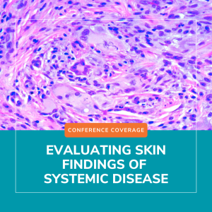
At the 2026 ODAC Dermatology Conference, attendees had the opportunity to learn about evaluating skin findings of systemic disease from Olayemi Sokumbi, MD, FAAD, professor of dermatology and laboratory medicine & pathology at Mayo Clinic. Dr. Sokumbi shared her expertise about evaluating skin findings that may occur in the context of systemic illness, emphasizing a structured derm-dermpath approach. Through two illustrative cases, she demonstrated how the skin serves as an early window to systemic disease and how clinicopathologic correlation (CPC) is integral to establishing the correct diagnosis.
Case 1:
Dr. Sokumbi described a gentleman in his 50s who presented with skin-colored papules on the ears accompanied by joint pains. The presence of concurrent systemic symptoms raised suspicion for an underlying systemic process, prompting a skin biopsy. Histopathology revealed foamy xanthomatous histiocytes, suggesting a non-Langerhans cell histiocytosis with inflammatory arthritis such as Erdheim-Chester disease (EDC). However, at least half of cases of EDC cases demonstrate a BRAF V600E mutation.1 Staining of this skin biopsy was negative for this mutation, prompting Dr. Sokumbi to return to the bedside and broaden the differential diagnosis. Subsequent physical examination revealed periungual papules and nodules, in a characteristic “coral beading” pattern, leading to the diagnosis of multicentric reticulohistiocytosis (MRH), a condition also associated with severe polyarthritis. The histopathologic pitfall requiring CPC was the presence of xanthomatized histiocytes, which are typical for EDC and underrecognized in MRH due to the rarity of this finding. Accurate diagnosis carries significant clinical implications, as MRH has a strong association with solid organ malignancy and requires therapeutic approaches distinct from those used in EDC.
Case 2:
Dr. Sokumbi presented the case of a young lady with diffuse cutaneous hyperpigmentation, which multiple providers had attributed to dermatoheliosis or photoaging. She highlighted, however, key photoprotected areas, such as the conchal bowls of the ears, also demonstrated blue-gray discoloration. The clinical differential diagnoses included lichen planus pigmentosus and argyria, yet the characteristic histopathologic features of these entities were not present on skin biopsy.
Instead, histologic examination revealed wavy deposits within the dermis that stained basophilic on Hematoxylin and Eosin and blue-black with Verhoeff Van Gieson (VVG) staining. Basophilic collagen fibers and altered deposits of elastic fibers have been reported as early-stage findings of ochronosis,3 in contrast to the classic late-stage yellow-brown banana-shaped collagen fibers. Based on these findings the patient was diagnosed with endogenous ochronosis/alkaptounuria, a genodermatosis characterized by impaired breakdown of tyrosine and phenylalanine.
In conclusion, Dr. Sokumbi emphasized how dermatologists are often uniquely positioned to diagnose systemic disease through careful evaluation of skin findings. Both cases underscored the importance of CPC. She encouraged repeating skin biopsies when the leading diagnosis remains unclear and collaborating with colleagues across specialties to ensure comprehensive management of systemic disease.
This session summary was written by Nagasai Adusumilli, MD, MBA, chief resident physician in dermatology at the George Washington University School of Medicine and Health Sciences.
References
- Haroche J, Cohen-Aubart F, Emile JF, et al. Reproducible and sustained efficacy of targeted therapy with vemurafenib in patients with BRAF(V600E)-mutated Erdheim-Chester disease. J Clin Oncol. 2015 Feb 10;33(5):411-8. PMID: 25422482.
- Camargo K, Pinkston O, Abril A, Sluzevich JC. Xanthomatous multicentric reticulohistiocytosis: an underrecognized variant. J Clin Rheumatol. 2018 Aug;24(5):285-287. PMID: 29239933.
- Chowdary S, Mahalingam M, Vashi NA. Reading between the layers: early histopathological findings in exogenous ochronosis. Am J Dermatopathol. 2014 Dec;36(12):989-91.PMID: 25415140.