Tag

dermoscopy

Practical Dermoscopy with Sima Jain, MD: In-depth Conference Coverage

By Medical Dermatology, ODAC Sessions No Comments
Dermoscopy tool image

Source: Next Steps in Derm

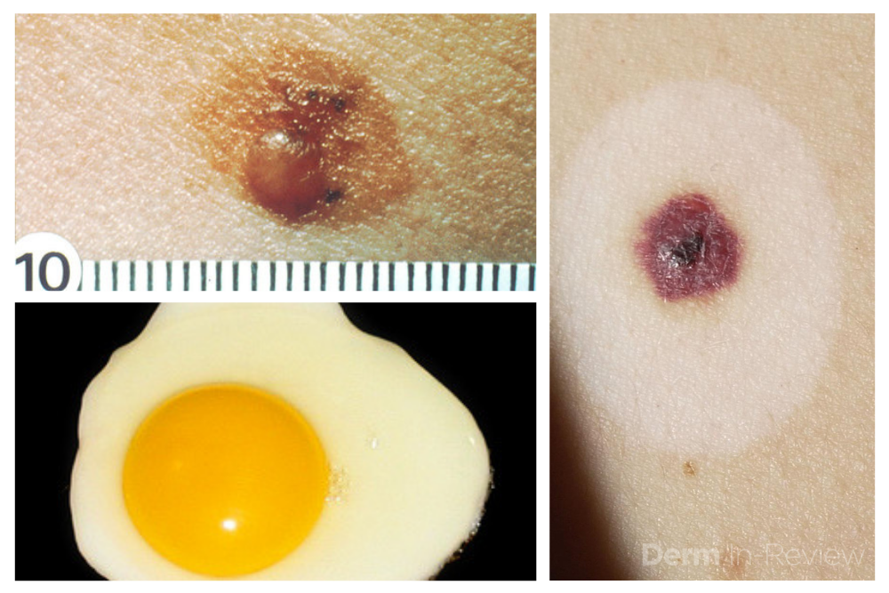
Dermoscopy, also known as epiluminescence microscopy, epiluminoscopy or skin surface microscopy, is an important way to visualize subsurface structures in the epidermis and dermis. In a 2-part series, Dr. Sima Jain reviews the evaluation of pigmented lesions, and the different vessel morphologies and patterns along with a discussion of specific findings in select cutaneous infections.

Read part 1 here

Read part 2 here

If you want to learn more about dermoscopy, make sure to register for the upcoming ODAC Dermatology, Aesthetic & Surgical Conference where Dr. Sima Jain will lead the following dermoscopy sessions:

The Utility of Dermoscopy in Challenging Clinical Cases

During this session, Dr. Jain will present challenging clinical cases and explore how dermoscopic evaluation can significantly increase clinical acumen.

Attendees will:

  1. Learn how to use dermoscopy to help differentiate types of alopecia
  2.  Learn how to use dermoscopic features to differentiate between melanoma and pigmented basal cell carcinoma
  3. Learn the dermoscopic features seen in cutaneous lupus and other rashes with follicular plugging.
PEARL ALERT!

Perifollicular scaling and fibrotic white dots are seen in lichen planopilaris. Orthogonal white streaks can be seen in melanoma. Glomerular vessels are often seen in Bowen’s disease.

Dermoscopy Essentials for Residents & Practicing Physicians

During this session, Dr. Jain will review the basics of dermoscopy and how it can be used to help diagnose both pigmented and non-pigmented skin lesions.

Attendees will:

  1. Learn how to correlate the colors seen on dermoscopic exam with histopathology.
  2. Learn how to distinguish non-melanocytic growths from melanocytic growths.
  3. Learn how to use vessel morphology to help diagnose cutaneous malignancies.
PEARL ALERT!

White streaks can be seen in melanoma and basal cell carcinoma. Telangiectasias, leaf-like areas and spoke wheel pigmentation are seen in basal cell carcinomas. The delta wing jet with contrail is specific for scabies.

Dr. Jean Bolognia’s Approach to Atypical Nevi

By Medical Dermatology, ODAC Sessions
Atypical Nevi on Patient leg

Source: Next Steps in Derm

This information was presented by Dr. Jean Bolognia at the 16th Annual ODAC Dermatology, Aesthetics and Surgical Conference held January 18th-21st, 2019 in Orlando, FL.  The highlights from her lecture were written and compiled by Dr. Daniel Yanes.

Despite being one of the more common reasons for consulting a dermatologist, the diagnosis and management of atypical nevi remain nuanced and can often be challenging. I had the opportunity to learn from Dr. Jean Bolognia on her approach to atypical nevi, and walked away with many pearls to share.

1. Identify the patient’s signature nevus and come up with a plan.

Sometimes it can be overwhelming to know where to begin when tasked with the patient who has numerous and atypical nevi. The first step is to identify the patient’s signature nevus. Do they tend to grow fried egg nevi, eclipse nevi, or cockade nevi? Are their signature moles all pink with little brown pigment, or are they pitch black with a wafer of scale? Identifying the signature nevus assists in determining the ugly duckling, and it will also help you develop a practical approach. In addition, if the patient has primarily pink nevi, palpation for induration versus soft flabbiness is helpful as banal intradermal melanocytic nevi can be pink in color.  If the patient has primarily small flat black nevi, you should hone in on the presence of inflammation that is not simply due to acne or folliculitis. Creating an individualized plan is the key to a successful examination.

2. Nevi change, and sometimes it is simply an aging phenomenon.

In addition to identifying the signature nevus, it is also essential to understand how melanocytic nevi evolve over time. While nevi classically progress from junctional to compound and then to dermal, sometimes they simply fade away. In the case of fried egg nevi, the “yolk” becomes more raised and softer over time while the “white” of the egg gradually fades (figure 1). This results in multiple large dermal nevi on the trunk in an older patient. Patients can be taught that when a nevus elevates, determining if the lesion is firm versus soft can assist in distinguishing between the need for evaluation versus an aging phenomenon. Although not all changing nevi are concerning nevi, it is still essential to give the patient’s nevus of concern special attention, even if it doesn’t catch your eye at first.

Continue reading.