Category

ODAC Sessions

Facial Anatomy: ODAC Coverage with Susan Weinkle, MD

By Aesthetic Dermatology, ODAC Sessions
Patient receiving injections

Source: Next Steps in Derm

This information was presented by Dr. Susan Weinkle at the 16th Annual ODAC Dermatology, Aesthetics and Surgical Conference held January 18th-21st, 2019 in Orlando, FL. The highlights from her lecture and live demonstrations were written and compiled by Dr Nikhil Shyam.

Dr. Susan Weinkle, an expert in the field of aesthetic and surgical dermatology, shares important anatomical concepts to consider when using neurotoxins and fillers safely.

The face can be split into 3 zones – the upper 1/3rd, the middle 1/3rd and the lower 1/3rd. Knowing the important vessels and nerves in each zone as well as their corresponding depth in the skin is crucial in order to minimize injury and utilize a safe technique when injecting neurotoxins and fillers.

Figure 1: D corresponds to areas where deeper injections are safer to avoid vasculature and nerves. S corresponds to areas where superficial (intradermal injections) are safer to avoid vasculature and nerves. Vessels shown are cartoon depictions of some of the more common arteries including the supraorbital, supratrochlear, superficial temporal, facial and superior and inferior labial arteries. Also depicted are the supraorbital, infraorbital and mental foramen that are located along the medial limbus.

Important Landmarks for the Upper 1/3rd of the Face

  • A line drawn vertically along the medial limbus denotes the anatomical locations of the supraorbital, infraorbital and mental foramen.
  • The supraorbital notch or foramen is the exit aperture for the supraorbital neurovascular bundle (NVB). While the vessels here initially lie deep in the skin, they become more superficial about 1.5 cm superior to the supraorbital foramen as they supply the forehead.
  • The supratrochlear NVB lies about 8-12 mm medial to the supraorbital NVB. The vessels are similarly deep initially and gradually become more superficial as they traverse superiorly to supply the forehead.
  • The supraorbital nerve, after exiting through its foramen, runs laterally and then superiorly about 1 cm medial to the temporal fusion line. It lies deep within the skin in this region.

Key Concepts for Upper 1/3rd Facial Injections and more.

Fine Tune Staging Risk for SCC

By Medical Dermatology, ODAC Sessions
Patel at ODAC Mohs

Source: Dermatology News

When caring for individuals with sun-damaged skin, dermatologists need comfort with the full spectrum of photo-related skin disease. From assessment and treatment of actinic keratoses (AKs) and field cancerization, to long-term follow-up of cutaneous squamous cell carcinomas (SCCs), appropriate treatment and staging can improve patient quality of life and reduce health care costs, Vishal Patel, MD, said at the Orlando Dermatology Aesthetic and Clinical Conference.

“Actinic keratosis/squamous cell carcinoma in situ is not a disease; it’s a symptom of cutaneous carcinogenesis or field cancerization,” said Dr. Patel, director of cutaneous oncology at George Washington University Cancer Center, Washington. On the other hand, he added, “field disease can be a marker for invasive squamous cell carcinoma risk, and it requires field treatment.” Treatment that reduces field disease is primary prevention because it decreases the formation of invasive SCC, he noted.

Read More….

Fungus Among US: Practical Case-Based Dermatophytosis

By Medical Dermatology, ODAC Sessions
Patient with fungus on foot

Source: Next Steps in Derm

This information was presented by Dr. Adam Friedman at the 16th Annual ODAC Dermatology, Aesthetics and Surgical Conference held January 18th-21st, 2019 in Orlando, FL.

Dermatophytosis constitutes a big chunk of “bread and butter” in dermatology.  In fact, an average of 4.1 million visits a year were due to dermatophytosis from 1995 to 2004! Nevertheless, these fungi can still stump the most seasoned dermatologist, and misdiagnosis can be surprisingly common. Dr. Adam Friedman, Professor, Interim Chair, and Program Director of Dermatology at George Washington School of Medicine and Health Sciences, recently presented interesting cases and practical pearls on how to diagnose and treat dermatophytosis. Here are some highlights.

Make the Diagnosis

Here’s the golden rule: if there is scale, scrape it! KOH preparation is first line in diagnosis of dermatophytosis.  Do you follow this rule? A recent survey showed that the percentages of dermatologists who scrape when suspicious of dermatophytosis were only 20-30% (always) and 30-40% (very often). Next, histology can be helpful in diagnosing nail fungus and Majocci’s granuloma (where KOH is usually negative).  Fungal culture be helpful to guide anti-fungal therapy, especially for tinea capitis in children. 

Tinea Pedis

Tinea pedis is the most common form of skin fungal infection, and there are 4 types: moccasin, interdigital, bullous, and ulcerative.

Don’t forget that non-dermatophytes (S. dimidiatum; S. hyalinum) can cause identical findings!  Also, an exuberant dermatophytid (or “id”) reaction, an inflammatory response to the fungal infection, can accompany findings of dermatophytosis. When you see a 2-hand-1-foot (or vice-versa) involvement, this can be another clue for diagnosing tinea pedis.

While topical azoles (econazole, other azoles) and allylamines (terbinafine, naftifine) and antifungal powder/spray weekly to shoes have been the mainstay treatment, there are some new topical options available.  Luliconazole 1% cream (daily for 2 week) for moist macerated web space; naftifine 2% gel and cream (daily for 2 week) for dry, scaling plaques; and urea 40% cream for moccasin tinea pedis have shown efficacy.

What about systemic anti-fungal therapy? The moccasin type and vesicular type may warrant oral terbinafine 250mg BID for 2-6 weeks and 2 weeks, respectively.  Since the vesicular type may have superimposed bacterial infection, an oral antibiotic may also be considered.

For more Tinea, click here.

ODAC and JDD Award Dr. Alan Menter

By Medical Dermatology, ODAC Sessions, Patient Care
Dr. Mentor Presenting at ODAC

Source: Practical Dermatology

Alan Menter, MD, has been awarded the Outstanding Researcher and Educator in Psoriatic Disease Award by the ODAC Dermatology, Aesthetics & Surgical Conference, in partnership with the Journal of Drugs in Dermatology (JDD).

The award recognizes Dr. Menter’s significant contribution and lifetime commitment to the advancement of psoriatic disease research as well as his work guiding the next generation of psoriasis experts and researchers.

“Dr. Menter has dedicated his career to improving psoriasis treatment options and standards of care while also pouring countless hours into up-and-coming psoriasis experts and researchers, ensuring his legacy will continue for generations to come,” said Shelley Tanner, CEO and president of SanovaWorks, which produces the JDD and ODAC.

Read More….

Male vs. Female Aesthetic Consultation – ODAC Dermatology Conference

By Aesthetic Dermatology, ODAC Sessions, Patient Care
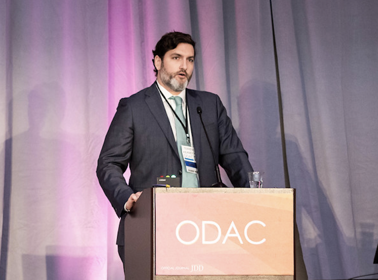
Terrence Keaney Male Aesthetics at ODAC

Source: Dermatology News

The first time a man walks in for an aesthetic consultation, he probably doesn’t know what to expect, according to Terrence Keaney, MD, a dermatologist in private practice in Arlington, Va. And he may not even be sure what he’s looking for.

However, he probably knows what he doesn’t like about his appearance. When men are questioned, the three areas they are most concerned about is their hairline, their eyes, and their jawline, said Dr. Keaney, speaking at the Orlando Dermatology Aesthetic and Clinical Conference.

It’s important to evaluate men differently, not just for anatomic differences from women, but also for behavioral and psychological factors unique to men as aesthetic patients, he noted.

Hot Topics in Infectious Disease

By Medical Dermatology, ODAC Sessions
Justin Finch MD Presenting at ODAC Dermatology Conference

Source: Dermatology News

New tricks from ticks, near-zero Zika, and the perils of personal grooming: Dermatologists have a lot to think about along the infectious disease spectrum in 2019, according to Justin Finch, MD, speaking at the Orlando Dermatology Aesthetic and Clinical Conference.

Anaphylaxis from alpha-gal syndrome is on the rise, caused in part by the geographic spread of the Lone Star tick. Beginning in 2006, isolated cases of an anaphylactic reaction to cetuximab, the epidermal growth factor receptor antagonist used to treat certain cancers, began to be seen in a curious geographic distribution. “The anaphylaxis cases were restricted to the southeastern United States, the home of the Lone Star tick,” said Dr. Finch, of the department of dermatology at the University of Connecticut, Farmington.

With some detective work, physicians and epidemiologists eventually determined that patients were reacting to an oligosaccharide called galactose-alpha–1,3-galactose (alpha-gal) found in cetuximab. This protein is also found in the meat of nonprimate mammals; individuals in the southeastern United States, where the Lone Star tick is endemic, had been sensitized via exposure to alpha-gal from Lone Star tick bites.

“Alpha-gal syndrome is on the rise,” said Dr. Finch, driven by the increased spread of this tick. Individuals who are sensitized develop delayed anaphylaxis 2-7 hours after ingesting red meat such as beef, pork, or lamb. “Ask about it,” said Dr. Finch, in patients who develop urticaria, dyspnea, angioedema, or hypotension without a clear offender. Because of the delay between allergen ingestion and anaphylaxis, it can be hard to connect the dots.


Hyperhydrosis: Where Are We Now?

By Medical Dermatology, ODAC Sessions
Video of Dr. Adam Friedman

Source:Dermatology News

When you extend your hand to a new patient, and he reflexively wipes his palm before shaking hands, be alert. It’s possible you’re seeing primary hyperhidrosis, a condition that’s both more common and more disabling than once thought.

“Looking at the biology of sweating, normally, it’s a good thing – we need it to survive. However, hyperhidrosis is too much of a good thing – it’s an excess of what is needed for normal biology,” said Adam Friedman, MD, speaking at the Orlando Dermatology Aesthetic and Clinical Conference.

Recent data, he pointed out, show that hyperhidrosis is more prevalent than previously thought – about 4.8% of individuals may have the condition, with about half having axillary hyperhidrosis. Symptoms peak in early adulthood, with adults aged 18-54 most affected. “These are the prime working years,” he said.

About 2% of teens are affected, and many adults report that symptoms began before they were 12 years old. Hand hyperhidrosis is a factor for computer and electronic device work, sports, and even handling paper and pencils, noted Dr. Friedman, professor of dermatology at George Washington University, Washington.

“Does it affect quality of life? Yes. We have data to support the impact. The adverse impact is actually greater than that of eczema and psoriasis,” he said, adding that patients won’t always bring up their concerns about sweating. “Often, it’s the patient who apologizes for having sweaty palms or who sticks to the paper on the exam table. It’s worth asking these patients if they are bothered by excessive sweating.”

Read More….

ODAC Dermatology Conference Returns to Orlando, Florida

By Aesthetic Dermatology, Medical Dermatology, ODAC Sessions, Patient Care, Surgical Dermatology
Orlando Florida Hotel

Source: Dermatology Times

The Orlando Dermatology Aesthetic & Clinical Conference (ODAC), formerly known as Orlando Derm, is scheduled for January 18-21 at the JW Marriott in Orlando.

This year’s meeting will open with presentations from physicians who will address advances in treating skin of color, hot topics in surgical dermatology and cutaneous malignancy, the latest on photodynamic therapy, and a year in review from the Journal of Drugs in Dermatology, among others.

Drs. Eric Bernstein and Jason Pozner will host a panel discussion on “My Top Picks for Laser and Energy Based Treatments.” And, Dr. Joel Cohen will give an overview of facial arterial supply.

During the general session on Saturday, Jan. 19, Dr. Brian Berman will address managing urticaria, which will be followed by talks by Dr. Deirdre Hooper on platelet rich plasma for hair growth and skin rejuvenation; Dr. Andrew Alexis on keloids and disorders of hyperpigmentation in skin of color; and, Drs. Bernstein and Pozner will address advances in non-surgical skin tightening.

On Sunday, January 20, Dr. Jean Bolognia will open the day’s general session with a review of advances in systemic therapies for melanoma.

For more information, visit ODAC online at https://orlandoderm.org.

Vascular Compromise

By ODAC Sessions, Patient Care
Dr. Joel Cohen presenting at ODAC dermatology conference

Source: Dermatology Times

Avoiding and treating vascular compromise with hyaluronic acid (HA) injections requires understanding the subtleties of underlying facial anatomy and keeping a well-stocked arsenal of treatments for impending necrosis, said an expert at the Orlando Dermatology Aesthetic and Clinical Conference (ODAC) in Miami.

“Some blood vessels may actually be in different locations than in some of the anatomic diagrams and cartoons that have long characterized their course,” said vice conference chair Joel L. Cohen, M.D., from Greenwood Village, Colorado, who serves on the teaching faculty for both the University of California, Irvine and the University of Colorado.

Although textbooks commonly depict the angular artery tracking adjacent to the nasofacial sulcus, he said, “it’s more common for the angular artery to be more lateral to that area, closer to the infraorbital distribution. In a recent cadaver study, only 19% of the time did the facial artery actually project upward along the side of the nose-cheek junction. But 32% of the time, the angular artery came off the facial artery earlier, and therefore coursed to more of the medial cheek area.”

The technique of aspirating before injecting is not foolproof. “There can be false negatives. A study indicates you probably have to pull back on the plunger for several seconds in order to physically be able to see if you’re in a vessel. We all surely realize that it is very difficult to have the needle in the exact spot you plan to inject, and then reposition your hand to pull back on the plunger of the syringe to try to aspirate, and then have your needle-tip remain in the exact same spot when you reposition again in order to inject. As you change your hand position to pull back on the plunger, you probably move a bit, maybe just a millimeter, from the original location to the location you later inject,” Dr. Cohen said.

A recent report also shows that it is possible to puncture and get into a vessel with small cannulas — the injectors aspirated blood despite using a cannula.

Read More…..

Five Pearls Target Wound Healing

By Medical Dermatology, ODAC Sessions, Patient Care, Surgical Dermatology
Robert Kirsner at the ODAC Dermatology Conference

Source: Dermatology News

Another reason not to prescribe opioids for postoperative pain – besides potentially adding to the epidemic the nation – comes from evidence showing these agents can impair wound healing.

In addition, epidermal sutures to close dermatologic surgery sites may be unnecessary if deep suturing is done proficiently. These and other pearls to optimize wound closure were suggested by Robert S. Kirsner, MD, PhD, professor and chair of the department of dermatology and cutaneous surgery at the University of Miami.

Avoid opioids for postoperative pain

“We know the opioid epidemic is a big problem. An estimated 5-8 million Americans use them for chronic pain,” Dr. Kirsner said at the Orlando Dermatology Aesthetic and Clinical Conference. “And there has been a steady increase in the use of illicit and prescription opioids.”

Emerging evidence suggests opioids also impair wound healing (J Invest Dermatol. 2017;137:2646-9). This study of 715 patients with leg ulcers, for example, showed use of opioids the most strongly associated with nonhealing at 12 weeks. “We found if you took an opioid you were less likely to heal,” Dr. Kirsner said. They found opioids significantly impaired healing, even when the investigators controlled for ulcer area, duration, and patient gender.

Read More….