Tag

ODAC 2019

Dr. Jean Bolognia’s Approach to Atypical Nevi

By Medical Dermatology, ODAC Sessions
Atypical Nevi on Patient leg

Source: Next Steps in Derm

This information was presented by Dr. Jean Bolognia at the 16th Annual ODAC Dermatology, Aesthetics and Surgical Conference held January 18th-21st, 2019 in Orlando, FL.  The highlights from her lecture were written and compiled by Dr. Daniel Yanes.

Despite being one of the more common reasons for consulting a dermatologist, the diagnosis and management of atypical nevi remain nuanced and can often be challenging. I had the opportunity to learn from Dr. Jean Bolognia on her approach to atypical nevi, and walked away with many pearls to share.

1. Identify the patient’s signature nevus and come up with a plan.

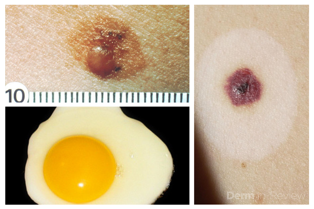
Sometimes it can be overwhelming to know where to begin when tasked with the patient who has numerous and atypical nevi. The first step is to identify the patient’s signature nevus. Do they tend to grow fried egg nevi, eclipse nevi, or cockade nevi? Are their signature moles all pink with little brown pigment, or are they pitch black with a wafer of scale? Identifying the signature nevus assists in determining the ugly duckling, and it will also help you develop a practical approach. In addition, if the patient has primarily pink nevi, palpation for induration versus soft flabbiness is helpful as banal intradermal melanocytic nevi can be pink in color.  If the patient has primarily small flat black nevi, you should hone in on the presence of inflammation that is not simply due to acne or folliculitis. Creating an individualized plan is the key to a successful examination.

2. Nevi change, and sometimes it is simply an aging phenomenon.

In addition to identifying the signature nevus, it is also essential to understand how melanocytic nevi evolve over time. While nevi classically progress from junctional to compound and then to dermal, sometimes they simply fade away. In the case of fried egg nevi, the “yolk” becomes more raised and softer over time while the “white” of the egg gradually fades (figure 1). This results in multiple large dermal nevi on the trunk in an older patient. Patients can be taught that when a nevus elevates, determining if the lesion is firm versus soft can assist in distinguishing between the need for evaluation versus an aging phenomenon. Although not all changing nevi are concerning nevi, it is still essential to give the patient’s nevus of concern special attention, even if it doesn’t catch your eye at first.

Continue reading. 

Systemic Therapies for Melanoma: What Every Dermatologist Needs to Know

By Medical Dermatology, ODAC Sessions
List of current melanoma therapies

Source: Next Steps in Derm

This information was presented by Dr. Jean Bolognia at the 16th Annual ODAC Dermatology, Aesthetics and Surgical Conference held January 18th-21st, 2019 in Orlando, FL.  The highlights from her lecture were written and compiled by Dr. Daniel Yanes, one of the 5 residents selected to participate in the Sun Resident Career Mentorship Program (a program supported by an educational grant from Sun Pharmaceutical Industries, Inc.). Dr. Yanes was paired with Dr. Jean Bolognia as his mentor.  

The world of melanoma is evolving, and dermatologists need to be equipped with the knowledge to help their patients navigate this landscape. Newer therapies for patients with more advanced stages of melanoma have not only drastically improved survival, but we as dermatologists must be prepared to recognize and treat the cutaneous side effects of these medications. This is a brief summary of common systemic therapies for melanoma with which every dermatologist should become familiar.

MAP Kinase Pathway Inhibitors

Selective BRAF Inhibitors

Melanoma tumor cells often have activating mutations that lead to constitutive activation of the MAP kinase pathway (See figure). Such activation can then lead to unregulated cell growth and proliferation. The most commonly detected mutation in BRAF results in the substitution of glutamic acid (E) for valine (V) at the 600th position in the BRAF protein and is referred to as BRAF V600E. Selective BRAF inhibitors, e.g. dabrafenib, encorafenib and vemurafenib, specifically target altered BRAF proteins. You can easily recognize these medications from their names, with raf indicating they target (B)RAF and nib identifying them as inhibitors. They are administered orally and chronically and lead to rapid responses but unfortunately tumor resistance commonly develops, often within six months. There are cutaneous side effects that the dermatologist should recognize, including morbilliform and folliculocentric eruptions, UVA photosensitivity (e.g. vemurafenib), keratoacanthomas/squamous cell carcinomas, and changes in melanocytic nevi (eruptive, enlargement, involution).

MEK Inhibitors

When mechanisms of resistance to selective BRAF inhibitors were investigated, a common finding was re-activation of the MAP kinase pathway via activation of MEK, another kinase that is downstream from BRAF. MEK inhibitors, e.g. binimetinib, cobimetinib, trametinib, were then combined with selective BRAF inhibitors to reduce the development of tumor resistance. These drugs are identified by the presence of a -metinib suffix. Interestingly, compared to BRAF inhibitors alone, combination BRAF+MEK therapy is associated with significantly less, not additive, cutaneous side effects – a real benefit to the patient.

Immunotherapy – Checkpoint Inhibitors

Immunotherapy is designed to stimulate the immune system to attack immunogenic melanoma cells. These monoclonal antibodies inhibit inhibitory signals that normally downregulate the immune system and thus act as immune checkpoints. These drugs model after the saying “the enemy of my enemy is my friend,” only it’s now “the inhibitor of the immune inhibitor is the immune stimulator.” CTLA4 is a receptor on regulatory T cells that plays an important role in diminishing immune responses. By blocking the inhibitory function of CTLA4 during the priming phase, the anti-CTLA4 antibody ipilimumab increases T cell immune activity. Peripherally, when the PD-1 receptor on T cells binds to its ligand, PD-L1, on tumor cells, an inhibitory signal results. In a similar fashion, the anti-PD-1 monoclonal antibodies approved for melanoma – nivolumab and pembrolizumab – can increase anti-tumor immune activity. Anti-PD-L1 monoclonal antibodies (e.g. avelumab, atezolizumab, durvalumab) have been approved to treat other malignancies, including Merkel cell carcinoma.

Read more. 

Dr. Jean Bolognia and the Many Faces of Lupus

By Medical Dermatology, ODAC Sessions
Dr. Jean Bolognia presenting at ODAC Dermatology Conference

Source: Next Steps in Derm

This information was presented by Dr. Jean Bolognia at the 16th Annual ODAC Dermatology, Aesthetics and Surgical Conference held January 18th-21st, 2019 in Orlando, FL.  The highlights from her lecture were written and compiled by Dr. Daniel Yanes.

Just as systemic lupus erythematosus (LE) can have protean systemic manifestations, cutaneous LE can present in many different ways. When confronted with the many faces of mucocutaneous LE, the following pearls can be valuable.

1. Be Aware of the SLICC Criteria

In 2012, the Systemic Lupus International Collaborating Clinics (SLICC) developed a set of clinical and immunologic criteria to assist in the diagnosis of systemic LE.

To view the original image full size image, click here.

To meet criteria for systemic LE, a patient must fulfill at least four criteria, with at least one clinical criterion and one immunological criterion OR have biopsy-proven lupus nephritis in the presence of ANA or anti-dsDNA antibodies. Of the 11 clinical criteria, 4 are mucocutaneous: (1) acute or subacute cutaneous LE; (2) chronic cutaneous lupus; (3) non-scarring alopecia; and (4) oral or nasal ulcers. Of note, the dermatologist doesn’t just establish the diagnosis of cutaneous LE, but can also determine the specific types of autoantibodies the patient is forming as well as if the patient has systemic involvement (e.g. hematologic or renal abnormalities). Remember the latter requires a urinalysis in addition to bloodwork. Acute cutaneous LE is more closely associated with systemic LE than subacute cutaneous LE, which in turn is more closely associated with systemic LE than discoid LE. The cutaneous manifestations of LE per the SLICC classification scheme are as follows:

Acute cutaneous lupus

  • Lupus malar rash (does not count if malar discoid)
  • Bullous eruption of systemic LE
  • Toxic epidermal necrolysis variant of systemic LE [also sometimes referred to as acute syndrome of apoptotic pan-epidermolysis (ASAP)]
  • Maculopapular lupus rash
  • Photosensitive lupus rash in the absence of dermatomyositis

Subacute cutaneous lupus

Chronic cutaneous lupus

  • Classic discoid LE
  • localized (above the neck)
  • disseminated (above and below the neck)
  • Hypertrophic (verrucous) LE
  • Mucosal LE
  • Lupus panniculitis (profundus)
  • LE tumidus
  • Chilblain lupus
  • Discoid LE/lichen planus overlap

2. A Positive ANA Does Not Equate to Systemic LE.

While ANA titer is positive in nearly all patients with systemic LE, not all patients with a positive ANA titer have LE.

Continue reading. 

Dermatology Contract Negotiations and Legal Tips

By ODAC Sessions, Patient Care
Dermatology Business deal

Source: Next Steps in Derm

If you are navigating the confusing landscape of contract negotiations or considering partnership in a private practice after years of employment, then take note! Ron Lebow, Senior Counsel in the Health Law practice group at Greenspoon Marder LLP, led a fantastic workshop at the 2019 ODAC conference. He covered common concerns when negotiating employment agreements with dermatology practices, as well as issues to address when it comes time to become a partner.

The most important point and recurring theme of Ron Lebow’s workshop was simply this:

“Make sure everything you want is in writing. Nothing is inferred. Everything is written and agreed upon by both parties.”

This point was stressed time and time again and will continue to be the overarching theme throughout the many parts of contract negotiations. With this in mind, here are some of the main takeaways and important questions we, dermatologists, should constantly keep in mind.

What Do I Do When Searching For A Job?

Surround yourself with experts! Find people who have your back. Make sure you find a lawyer who deals with medical professionals (particularly your specialty) for a living. You may need to find an accountant to work with you depending on your situation. Lastly, it may be in your best interest to work through recruiters. Some recruiters are better than others, and Ron Lebow recommends Dermatology Authority as an organization that provides good recruiting services for dermatologists.

Can I Negotiate When Looking For A Job?

When dealing with negotiations, it is important to recognize the many aspects of the process. With respect to the job you are offered:

  • They expect you to negotiate. It’s not rude to negotiate. It is part of the process.
  • They aren’t doing you a favor by hiring you. They are making a profit on you and therefore, you do have some leverage in the process. You can negotiate because they want you.
  • They will (almost) never take the offer away. The only instance in which they may take an offer away is if you ask for something, they say no, and then you continue to ask for something that is already off the table.
  • Don’t negotiate before you bring a lawyer in to help with the contract and negotiation. It may already be too late for them to be useful.
  • Don’t sign term sheet/offer letter until you have spoken with a lawyer who has reviewed the information. Again, it may already be too late for them to be useful.
  • The term of the contract, the length of time you sign for, is a non-issue. The real term of the contract is actually the notice you have to give before leaving, not the actual term of the contract itself. For example, if you sign a two-year contract but the notice you must give is three months, the term for all intents and purposes is three months from the time you decide you want to break your contact.
  • Make sure it is spelled out when partnership will be happening if that is on the table. Three years is a common timetable for partnership to be offered. If partnership is a possibility, make sure there is something written in the contract in case ownership changes between the time you start and the time you would have been allowed to start buying into partnership (e.g. if the group sells to private equity).

Compensation and Bonuses

Continue reading. 

Actinic Keratosis and Squamous Cell Carcinoma: Pearls from ODAC

By Medical Dermatology, ODAC Sessions
Dr. Patel presenting at ODAC Dermatology Conference

Source: Next Steps in Derm

In a 20-minute lecture presented at the 16th Annual ODAC conference, Dr. Patel reviewed the appropriate management of actinic keratoses and squamous cell carcinoma. Grabbing the attention of the audience early on, Dr. Patel quoted the staggering statistics for squamous cell carcinoma – calling the growing epidemic “a public health crisis.”  He challenged dermatologists to lead the charge in a more sophisticated approach to disease stratification.

Data are conflicting regarding the risk of progression of actinic keratoses to squamous cell carcinoma. Despite Dr. Patel’s expertise, he admitted even he finds it impossible to predict which lesions will progress. Instead, he takes a more astute approach and taking a step back to focus on the burden of disease.

Twenty is the magic number – over 20 actinic keratoses increase the risk of squamous cell carcinoma.

With the groundwork firmly laid, Dr. Patel delved into the crux of his talk. He posed a thought-provoking question to captivated listeners:  Are actinic keratoses a disease or a symptom? In the same way hypertension leads to stroke, actinic keratoses lead to squamous cell carcinoma.

Actinic keratoses are a field disease, as such we should focus on field treatment.

Dr. Patel drove his point home with several instructive clinical cases. With each patient, Dr. Patel calls dermatologists to first discern field disease from invasive disease. Once invasive disease has been excluded, hyperkeratotic lesions should be debrided, and a strict regimen of topical 5-fluorouracil instituted for 4 weeks. This regimen should be followed by photodynamic therapy in 3 months.

Continue reading. 

Facial Anatomy: ODAC Coverage with Susan Weinkle, MD

By Aesthetic Dermatology, ODAC Sessions
Patient receiving injections

Source: Next Steps in Derm

This information was presented by Dr. Susan Weinkle at the 16th Annual ODAC Dermatology, Aesthetics and Surgical Conference held January 18th-21st, 2019 in Orlando, FL. The highlights from her lecture and live demonstrations were written and compiled by Dr Nikhil Shyam.

Dr. Susan Weinkle, an expert in the field of aesthetic and surgical dermatology, shares important anatomical concepts to consider when using neurotoxins and fillers safely.

The face can be split into 3 zones – the upper 1/3rd, the middle 1/3rd and the lower 1/3rd. Knowing the important vessels and nerves in each zone as well as their corresponding depth in the skin is crucial in order to minimize injury and utilize a safe technique when injecting neurotoxins and fillers.

Figure 1: D corresponds to areas where deeper injections are safer to avoid vasculature and nerves. S corresponds to areas where superficial (intradermal injections) are safer to avoid vasculature and nerves. Vessels shown are cartoon depictions of some of the more common arteries including the supraorbital, supratrochlear, superficial temporal, facial and superior and inferior labial arteries. Also depicted are the supraorbital, infraorbital and mental foramen that are located along the medial limbus.

Important Landmarks for the Upper 1/3rd of the Face

  • A line drawn vertically along the medial limbus denotes the anatomical locations of the supraorbital, infraorbital and mental foramen.
  • The supraorbital notch or foramen is the exit aperture for the supraorbital neurovascular bundle (NVB). While the vessels here initially lie deep in the skin, they become more superficial about 1.5 cm superior to the supraorbital foramen as they supply the forehead.
  • The supratrochlear NVB lies about 8-12 mm medial to the supraorbital NVB. The vessels are similarly deep initially and gradually become more superficial as they traverse superiorly to supply the forehead.
  • The supraorbital nerve, after exiting through its foramen, runs laterally and then superiorly about 1 cm medial to the temporal fusion line. It lies deep within the skin in this region.

Key Concepts for Upper 1/3rd Facial Injections and more.

Fine Tune Staging Risk for SCC

By Medical Dermatology, ODAC Sessions
Patel at ODAC Mohs

Source: Dermatology News

When caring for individuals with sun-damaged skin, dermatologists need comfort with the full spectrum of photo-related skin disease. From assessment and treatment of actinic keratoses (AKs) and field cancerization, to long-term follow-up of cutaneous squamous cell carcinomas (SCCs), appropriate treatment and staging can improve patient quality of life and reduce health care costs, Vishal Patel, MD, said at the Orlando Dermatology Aesthetic and Clinical Conference.

“Actinic keratosis/squamous cell carcinoma in situ is not a disease; it’s a symptom of cutaneous carcinogenesis or field cancerization,” said Dr. Patel, director of cutaneous oncology at George Washington University Cancer Center, Washington. On the other hand, he added, “field disease can be a marker for invasive squamous cell carcinoma risk, and it requires field treatment.” Treatment that reduces field disease is primary prevention because it decreases the formation of invasive SCC, he noted.

Read More….

Fungus Among US: Practical Case-Based Dermatophytosis

By Medical Dermatology, ODAC Sessions
Patient with fungus on foot

Source: Next Steps in Derm

This information was presented by Dr. Adam Friedman at the 16th Annual ODAC Dermatology, Aesthetics and Surgical Conference held January 18th-21st, 2019 in Orlando, FL.

Dermatophytosis constitutes a big chunk of “bread and butter” in dermatology.  In fact, an average of 4.1 million visits a year were due to dermatophytosis from 1995 to 2004! Nevertheless, these fungi can still stump the most seasoned dermatologist, and misdiagnosis can be surprisingly common. Dr. Adam Friedman, Professor, Interim Chair, and Program Director of Dermatology at George Washington School of Medicine and Health Sciences, recently presented interesting cases and practical pearls on how to diagnose and treat dermatophytosis. Here are some highlights.

Make the Diagnosis

Here’s the golden rule: if there is scale, scrape it! KOH preparation is first line in diagnosis of dermatophytosis.  Do you follow this rule? A recent survey showed that the percentages of dermatologists who scrape when suspicious of dermatophytosis were only 20-30% (always) and 30-40% (very often). Next, histology can be helpful in diagnosing nail fungus and Majocci’s granuloma (where KOH is usually negative).  Fungal culture be helpful to guide anti-fungal therapy, especially for tinea capitis in children. 

Tinea Pedis

Tinea pedis is the most common form of skin fungal infection, and there are 4 types: moccasin, interdigital, bullous, and ulcerative.

Don’t forget that non-dermatophytes (S. dimidiatum; S. hyalinum) can cause identical findings!  Also, an exuberant dermatophytid (or “id”) reaction, an inflammatory response to the fungal infection, can accompany findings of dermatophytosis. When you see a 2-hand-1-foot (or vice-versa) involvement, this can be another clue for diagnosing tinea pedis.

While topical azoles (econazole, other azoles) and allylamines (terbinafine, naftifine) and antifungal powder/spray weekly to shoes have been the mainstay treatment, there are some new topical options available.  Luliconazole 1% cream (daily for 2 week) for moist macerated web space; naftifine 2% gel and cream (daily for 2 week) for dry, scaling plaques; and urea 40% cream for moccasin tinea pedis have shown efficacy.

What about systemic anti-fungal therapy? The moccasin type and vesicular type may warrant oral terbinafine 250mg BID for 2-6 weeks and 2 weeks, respectively.  Since the vesicular type may have superimposed bacterial infection, an oral antibiotic may also be considered.

For more Tinea, click here.

ODAC and JDD Award Dr. Alan Menter

By Medical Dermatology, ODAC Sessions, Patient Care
Dr. Mentor Presenting at ODAC

Source: Practical Dermatology

Alan Menter, MD, has been awarded the Outstanding Researcher and Educator in Psoriatic Disease Award by the ODAC Dermatology, Aesthetics & Surgical Conference, in partnership with the Journal of Drugs in Dermatology (JDD).

The award recognizes Dr. Menter’s significant contribution and lifetime commitment to the advancement of psoriatic disease research as well as his work guiding the next generation of psoriasis experts and researchers.

“Dr. Menter has dedicated his career to improving psoriasis treatment options and standards of care while also pouring countless hours into up-and-coming psoriasis experts and researchers, ensuring his legacy will continue for generations to come,” said Shelley Tanner, CEO and president of SanovaWorks, which produces the JDD and ODAC.

Read More….

Male vs. Female Aesthetic Consultation – ODAC Dermatology Conference

By Aesthetic Dermatology, ODAC Sessions, Patient Care
Terrence Keaney Male Aesthetics at ODAC

Source: Dermatology News

The first time a man walks in for an aesthetic consultation, he probably doesn’t know what to expect, according to Terrence Keaney, MD, a dermatologist in private practice in Arlington, Va. And he may not even be sure what he’s looking for.

However, he probably knows what he doesn’t like about his appearance. When men are questioned, the three areas they are most concerned about is their hairline, their eyes, and their jawline, said Dr. Keaney, speaking at the Orlando Dermatology Aesthetic and Clinical Conference.

It’s important to evaluate men differently, not just for anatomic differences from women, but also for behavioral and psychological factors unique to men as aesthetic patients, he noted.